047 月/24

KMD Bioscience-Immunoprecipitation: Applications and Protocols

Immunoprecipitation (IP) is a widely employed technique in molecular biology and biochemistry, enabling the isolation and concentration of specific proteins from complex mixtures. By leveraging the specificity of antibodies to selectively bind proteins, IP facilitates the study of protein-protein interactions, protein modifications, and protein functions. This article delves into the myriad applications of immunoprecipitation and outlines some common protocols utilized in this technique.

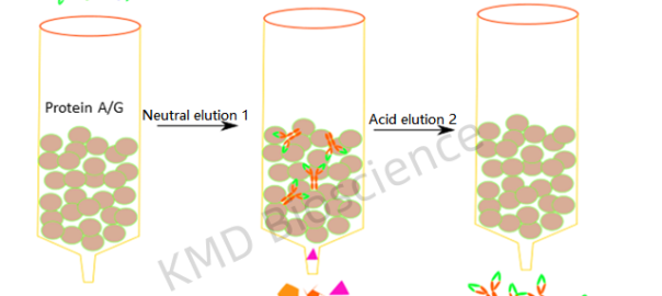
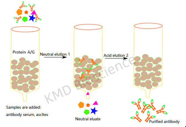
The essence of immunoprecipitation lies in the interaction between the antigen (target protein) and the antibody. A suitable antibody is chosen to selectively bind the protein of interest, forming an antigen-antibody complex. This complex is then precipitated from the solution, typically using protein A/G agarose beads. The beads are collected, and washed to remove unbound materials, and the bound proteins are eluted for further analysis. Understanding the core principles and methodology of IP is pivotal for obtaining reliable and interpretable results.

Immunoprecipitation (IP) and other protein interaction applications such as yeast two-hybrid (Y2H), protein microarrays, and Förster Resonance Energy Transfer (FRET) serve the common goal of elucidating protein interactions, albeit through distinct mechanisms. IP leverages the specificity of antibodies to isolate and concentrate particular proteins from a mixture, providing a direct method of assessing protein interactions or modifications in a near-native state. On the other hand, Y2H employs a genetic system in yeast to detect interactions, offering a high-throughput albeit less direct method, which may miss interactions occurring outside the yeast’s nuclear environment. Protein microarrays provide a high-throughput platform to study protein-protein interactions on a large scale, yet may not always reflect the natural cellular context. FRET utilizes energy transfer between two light-sensitive molecules to gauge the interaction and distance between proteins, offering real-time interaction analysis within living cells. Each of these methods has its own set of advantages and limitations concerning sensitivity, throughput, and contextual relevance, choosing method contingent on the specific requirements of the research question at hand.

Immunoprecipitation finds extensive applications in research fields like oncology, immunology, and neurobiology. It is instrumental in studying protein-protein interactions, identifying post-translational modifications, and investigating cellular signaling pathways. Moreover, IP serves as a crucial step in various downstream applications such as Western blotting, mass spectrometry, and enzyme activity assays which provide insights into the functional and structural aspects of proteins.

Here’s a brief protocol of Immunoprecipitation:

Step 1 Sample Preparation

Preparing a clear, homogenous sample is the first step, which involves lysing cells or tissues to release proteins. Adding protease and phosphatase inhibitors to prevent protein degradation.

Step 2 Antibody Selection and Binding

Choosing a high-affinity, specific antibody is crucial. The antibody is incubated with the sample to allow binding to the target protein. Incubate at an appropriate temperature for a specified time (e.g., 1-2 hours) to allow antibody-protein binding.

Step 3: Immobilization to a Solid Support

The antibody-protein complexes are then immobilized on a solid support, usually beads coated with protein A or G which bind the Fc region of antibodies. During this incubation, the antibody binds to its target protein, and the protein-bead-antibody complex forms.

Step 4 Washing and Elution

Washing the beads several times with the wash buffer to remove unbound proteins and contaminants, and the target protein is eluted from the beads, often by altering the pH or using a competitive ligand.

Step 5 Analysis

The eluted protein can be analyzed using various techniques like Western blotting, mass spectrometry, or gel electrophoresis.

Step 6 Optimization

Fine-tuning parameters such as incubation times, temperatures, and buffer compositions is often necessary to achieve optimal results.

Figure 1Immunoprecipitation. (A) Before an immunoprecipitation procedure begins, a protein of interest exists in a heterogenous sample with many other proteins. (B) A scientist adds antibodies that recognize the protein to small beads and then adds the beads to the sample. (C) The antibodies bind to the proteins, causing them to precipitate out of solution and adhere to the beads. (D) The beads are removed from the sample, purifying the protein of interest. The proteins can be removed from the beads by changing the salinity or pH of the solution.(Figure source: Matt Carter, Jennifer C. Shieh, 2010)


Immunoprecipitation remains a cornerstone technique, bridging the molecular interactions with the vast expanse of research insights. The meticulous adherence to protocols coupled with a profound understanding of its applications propels the scientific inquiry forward, opening avenues for novel discoveries and a deeper understanding of life’s molecular machinery.


KMD Bioscience has been dedicated to the study of protein-nucleic acid interactions for over 10 years. Eukaryotic genomic DNA exists in the form of chromatin, and the study of protein-DNA interactions in the chromatin environment is a fundamental way to elucidate the mechanisms of eukaryotic gene expression. Scientists in KMD Bioscience, which have accumulated a wealth of experience in immunoassay technology, are able to quickly customize protein-nucleic acid interaction study assays for different clients. With years of technical expertise, KMD Bioscience is not only able to complete the experimental content quickly, but also to ensure that each step of the experiment is carefully controlled to ensure that the test results are accurate, objective and credible. KMD Bioscience, which has complete protein detection equipment, has established mature and perfect antibody platforms, protein platforms and other technology platforms.

For more information, Visit us at  https://www.kmdbioscience.com/ to have a detailed understanding.

037 月/24

KMD Bioscience-Antibody Labeling and Conjugation Techniques

Antibodies are invaluable tools in the realms of biology and medicine, acting as precise detectives that identify specific targets within a complex milieu. However, their detective work is only visible to researchers when antibodies are tagged with detectable labels, a process known as antibody labeling or conjugation. The fundamental structure of an antibody is often likened to a Y-shaped figure, comprising four polypeptide chains – two identical heavy (H) chains and two identical light (L) chains. Heavy Chains: The heavy chains are longer polypeptide chains that form the base and part of the arms of the Y-shaped antibody. They are crucial for defining the class or isotype of the antibody. Light Chains: The light chains are shorter and align parallelly with the heavy chains, contributing to the formation of the arms of the antibody. This article elucidates the diverse techniques used for antibody labeling and conjugation, each catering to distinct research requirements and applications.

Direct Labeling

Fluorophore Conjugation

Directly conjugating fluorophores to antibodies allows for fluorescence-based detection. This is crucial in flow cytometry, immunofluorescence microscopy, and fluorescence-linked immunosorbent assay (FLISA). The marriage of fluorophores to antibodies via conjugation transforms the invisible detective work of antibodies into a visual spectacle, enabling researchers to track and analyze molecular and cellular events. The process of fluorophore conjugation to antibodies is a meticulous one, underscoring a fine balance between achieving detectable signals and preserving antibody functionality.

Enzyme Conjugation

Enzymes like horseradish peroxidase (HRP) or alkaline phosphatase (AP) are conjugated to antibodies for colorimetric detection. Widely used in enzyme-linked immunosorbent assay (ELISA) and Western blotting, the enzymatic reaction produces a color change indicating the presence of the target.

Radioisotope Conjugation

Antibodies tagged with radioactive isotopes enable radioimmunoassay (RIA), a sensitive technique for quantifying antigens.

Biotinylation

Conjugating biotin to antibodies facilitates high-affinity binding to streptavidin, allowing for versatile detection strategies.

Indirect Labeling

Secondary Antibody Conjugation

Here, a labeled secondary antibody, which is directed against the Fc region of the primary antibody, provides the detectable signal. This method amplifies the signal but may also increase background noise.

Tandem Labeling

Combining direct and indirect labeling, tandem conjugates utilize a secondary antibody conjugated to a large, highly fluorescent protein or polymer, enhancing signal intensity.

Genetic Fusion

Genetic techniques enable the fusion of antibodies with fluorescent proteins or enzymes at the DNA level, allowing for precise control over the labeling site.

Cysteine or Lysine-directed Conjugation

Chemical conjugation at specific amino acid residues like cysteine or lysine ensures a defined attachment site, reducing heterogeneity in the conjugate population.

Applications

Diagnostic Assays

Labeled antibodies are essential in diagnostic assays like ELISA, Western blotting, and RIA, enabling the detection and quantification of disease markers.

Imaging

In molecular imaging, labeled antibodies illuminate target structures, elucidating cellular architecture and processes.

Therapeutics

Antibody-drug conjugates (ADCs) are a burgeoning therapeutic modality, where labeled antibodies deliver cytotoxic drugs specifically to cancer cells.

Conclusion: The realm of antibody labeling and conjugation is a vibrant field, continuously evolving with emerging technologies. Each conjugation technique opens new vistas, enhancing the sensitivity, specificity, and multiplexing capabilities in research. The magic of labeled antibodies continues to unveil the intricacies of biology, driving forward the wheels of discovery, diagnostics, and therapeutics.

Antibody labeling and conjugation significantly expand the utility of antibodies across a wide spectrum of scientific and clinical arenas. Labeled antibodies are widely used in many applications: Immunofluorescence, Flow Cytometry, Western Blotting, Enzyme-Linked Immunosorbent Assay (ELISA), Immunohistochemistry (IHC) and Immunoprecipitation, etc.

KMD Bioscience offers a variety of high-quality labeling services to meet your specific needs. Antibodies or Proteins can both be cross-linked using different reporter enzymes such as horseradish phosphatase and alkaline phosphatase (AP). This allows for maximum retention of labeled antibody activity and chemical coupling. Enzyme-labeled proteins and antibodies can be used in ELISA, immunoblotting, in situ fusion, cell, and histochemical assays.

For more information, Visit us at https://www.kmdbioscience.com/ to have a detailed understanding.

References

1 Hermanson, G. T. (2013). Bioconjugate Techniques. Academic Press.

Junutula, J. R., Raab, H., Clark, S., Bhakta, S., Leipold, D. D., Weir, S., … & Liu, B. (2008). Site-specific conjugation of a cytotoxic drug to an antibody improves the therapeutic index. Nature Biotechnology, 26(8), 925-932.

2 Smith, M. E. B., Schumacher, F. F., Ryan, C. P., Tedaldi, L. M., Papaioannou, D., Waksman, G., … & Baker, J. R. (2010). Protein modification, bioconjugation, and disulfide bridging using bromomaleimides. Journal of the American Chemical Society, 132(6), 1960-1965.

3 Ackerman, M. E., & Chalouni, C. (2018). Antibody–Drug Conjugates for Cancer Therapy. Annual Review of Medicine, 69, 275-288.

027 月/24

KMD Bioscience-Antibody Purification Strategies: Affinity vs. Protein A/G

In the realm of biotechnology, antibodies are lauded as precision tools, driving insights in research, diagnostics, and therapeutics. Central to maximizing the potential of antibodies is the purification process, which ensures specificity, functionality, and purity. Two prevalent strategies in antibody purification are Affinity Purification and Protein A/G Purification. This article delves into the nuances of these techniques, drawing a comparative analysis to guide informed decision-making in antibody purification endeavors.


Affinity purification leverages the specific interaction between the antibody and an antigen or a specific ligand. The target antibody is captured onto a solid support to which the ligand is immobilized, facilitating purification from complex mixtures. The ligands can be tailored to the antibody of interest, providing a high degree of specificity and purity.

Affinity purification can be employed for different types of antibodies and is not confined to IgG antibodies alone.

Gentle elution conditions can be used during affinity purification, preserving the antibody’s functionality and integrity.

Fig. 1 Protein A / G affinity Purification

Protein A/G purification exploits the interaction between Protein A or Protein G and the Fc region of IgG antibodies. The Protein A or G is immobilized on a solid support, capturing the antibodies from the mixture. Protein A/G purification is amenable to a wide range of IgG antibodies from various species, making it a versatile choice. The robust interaction between Protein A/G and IgG antibodies often results in high purity levels, like 95%, 98% purity, making it a preferred choice for many applications. The elution conditions of protein A/G purification, typically involving a low pH, may potentially affect antibody stability and should be considered in the purification strategy.

Affinity purification, with its customizable ligand approach, may offer higher specificity, especially for non-IgG antibodies or subclass-specific purification. Protein A/G purification stands out for its broad-spectrum applicability to IgG antibodies across various species.

Both affinity purification and protein A/G methods can achieve high purity, but the yield may vary based on the affinity of the interactions and the specific protocol employed.

Protein A/G purification kits are readily available and can be more time and cost-effective, whereas affinity purification may require additional steps in ligand preparation and optimization.

In summary, the choice between affinity purification and Protein A/G purification depends on several factors, including the antibody’s characteristics, the desired purity level, the scale of purification, and the ease of use. The journey from a biological sample to a purified antibody is a meticulous expedition. Both Affinity Purification and Protein A/G Purification offer unique advantages, and the choice between them hinges on the specifics of the antibody in question, the downstream application, and the resources at hand. As science advances, so does the toolkit for antibody purification, each technique refined, and sometimes combined, to meet the evolving demands of antibody-based endeavors. The comparative insights drawn between Affinity and Protein A/G purification serve as a compass, guiding researchers through the cascade of antibody purification towards their quest for precision and purity.

KMD Bioscience is a professional provider of antibody and recombinant protein products and services, with years of pre-clinical research experience and production experience to provide upstream and downstream services for antigen and antibody production. Whether you have a clearly defined protocol or you just have an idea for an experiment, KMD Bioscience can tailor it to your needs. KMD Bioscience started from the phage display technology platform to provide high-quality antibody discovery services, and gradually established stable cell line construction and screening, natural protein extraction and fermentation, and recombinant protein custom expression services, a multi-species antibody discovery platform.

For more information, Visit us at https://www.kmdbioscience.com/ to have a detailed understanding.

017 月/24

KMD Bioscience-Flow Cytometry for Antibody

Flow Cytometry, a powerful tool at the crossroads of optical and cell sciences, provides a window into cell populations’ complex dynamics. At the heart of this technology lies the synergy between antibodies and fluorescent dyes, illuminating specific cellular constituents to paint a vivid picture of cellular heterogeneity, function, and health. This article journeys through the mechanics and applications of flow cytometry in antibody-based cell analysis, showcasing its indispensability in modern biological research. Flow cytometry leverages the principle of light scattering and fluorescence to analyze individual cells as they flow past laser beams. The interactions between light and cells, augmented by antibody-fluorophore conjugates, generate data that unveil the cells’ physical and molecular characteristics.

Critical to the success of flow cytometry is the conjugation of antibodies to fluorophores, which enables the specific detection of cellular markers. This labeling transforms the invisible molecular landscape of cells into a detectable, quantifiable entity. Modern flow cytometers have multiple lasers and detectors, enabling multi-parameter analysis. Using a panel of differently labeled antibodies, researchers can simultaneously probe multiple markers on or within cells, catapulting the depth and breadth of analysis.

Fig. 1 Cell Flow Cytometry Process


As we all know, FCM is widely used in cell analysis, here’s a draft conclusion of several applications:

Immunophenotyping

Flow cytometry shines in immunophenotyping, where it delineates the diverse immune cell subsets within a sample based on the expression of surface and intracellular markers. This is pivotal in immunology research, clinical diagnostics, and monitoring treatment responses.

Cell Cycle and Proliferation Analysis

Through antibodies targeting specific cell cycle markers, flow cytometry provides insight into cell cycle distribution and proliferation status, crucial in cancer research and developmental biology. Flow cytometry can be used to determine the distribution of cells in different phases of the cell cycle (G1, S, G2, and M phases). This is valuable for studying cell proliferation and cell cycle regulation.

Functional Assays

Antibody-based flow cytometry assays delve into cellular functions such as cytokine production, calcium flux, and apoptosis. These assays are instrumental in understanding cellular responses in health and disease.

Sorting and Isolation

Flow cytometry extends to cell sorting, where labeled cells are sorted based on specific markers, enabling the isolation of pure cell populations for further analysis or culture.

Monitoring Cell Health

Antibodies targeting markers of health and stress allow for the assessment of cell viability, activation status, and overall cellular health.

Clinical Diagnostics and Monitoring

In the clinical realm, flow cytometry aids in diagnosing and monitoring various conditions, including hematological malignancies and immune disorders.

Flow cytometry continues to evolve with advancements in technology and reagents. The advent of spectral flow cytometry, mass cytometry, and an expanding repertoire of antibody-fluorophore conjugates pushes the boundaries of what can be analyzed and understood about cellular populations. Flow cytometry, with its ability to dissect the cellular milieu in remarkable detail, remains a linchpin in antibody-based cell analysis. As researchers continue to harness and expand upon this technology, the insights gleaned edge us closer to a profound understanding of the cellular cosmos, with repercussions spanning basic research to clinical interventions. The interplay between antibodies and flow cytometry epitomizes a harmonious blend of specificity and analysis, propelling the exploration of the cellular frontier toward new horizons.


KMD Bioscience has been committed to cell biology technology research for many years and has experienced experimental technicians. KMD Bioscience has been engaged in flow cytometry operation for many years, and has accumulated rich experience in cell operation, mature and stable technology, and standardized operation process, to ensure to provide customers with real and reliable results. With advanced technical equipment and a complete cell culture platform, using advanced flow cytometers and other precision instruments, combined with specific detection reagents, KMD Bioscience can enable customers to get satisfactory cell flow cytometry reports in a short time and experience high-quality cell flow cytometry services. Other than that KMD Bioscience has a variety of data processing and analysis systems, which can provide customers with one-stop technical services from cell processing, on-machine detection, and data analysis, and can also provide after-sales technical consultation to solve customer problems and meet customer needs.

266 月/24

KMD Bioscience-Antibody Phage Display: A Revolutionary Approach

In the expansive landscape of antibody engineering and discovery, Antibody Phage Display emerges as a pivotal technology, marrying the specificity of antibodies with the versatility of phage display. Antibody phage display is a powerful technique used in molecular biology and biotechnology to generate and select monoclonal antibodies with high specificity and affinity for a particular target molecule, such as a protein, peptide, or small molecule. The technique is based on the use of bacteriophages (phages), which are viruses that infect bacteria. This innovative approach has been instrumental in advancing both fundamental research and therapeutic interventions. This article explores the genesis, mechanics, and impact of Antibody Phage Display, underscoring its transformative potential in the realm of modern biotechnology.

The inception of phage display in the late 1980s by George Smith provided a robust platform for peptide and protein expression on bacteriophage surfaces. The integration of antibody fragment expression into this system heralded the advent of Antibody Phage Display. The principle behind Antibody Phage Display is the expression of antibody fragments on the surface of bacteriophages, coupled with the encoding DNA within the phage. This genetic linkage between phenotype and genotype facilitates the high-throughput screening and selection of antibodies with desired specificities.

Fig. 1 Diagram of phage display technology

Mechanics of Antibody Phage Display

Library Construction

The journey begins with the construction of phage display libraries, encompassing a vast diversity of antibody fragments generated from immunized animals, humans, or synthetic sources. Antibody phage display begins with the creation of a diverse library of antibody fragments. This library typically consists of a large number of genetically engineered bacteriophages, each displaying a unique antibody fragment on its surface. These antibody fragments are usually single-chain variable fragments (scFv) or fragment antigen-binding (Fab) regions.

Biopanning

The library is then exposed to the target molecule (antigen) of interest. Phages displaying antibody fragments that bind to the antigen with high affinity are selectively retained, while non-binding or weakly binding phages are washed away. The crux of the technology is biopanning, a process of iterative rounds of selection under increasingly stringent conditions to enrich for phages displaying antibodies with high affinity and specificity towards the target antigen.

Expression and Characterization:

After several rounds of selection, the DNA encoding the antibody fragments from the enriched phage population is extracted and cloned. Post-selection, the identified antibody fragments are often expressed in suitable host cells, followed by a thorough characterization to assess their binding kinetics, specificity, and other functional attributes.

Applications and Impact

Therapeutic Antibody Discovery

Antibody Phage Display has been instrumental in the discovery of therapeutic antibodies, with notable examples like adalimumab (Humira®), the first fully human antibody generated via phage display. Antibody phage display is widely used in the development of therapeutic monoclonal antibodies for various diseases, including cancer, autoimmune disorders, and infectious diseases.

Biomarker Discovery

The technology aids in unraveling novel biomarkers by identifying antibodies against hitherto unknown cellular antigens, paving the way for diagnostic advancements.

Epitope Mapping

The precision of Antibody Phage Display facilitates fine mapping of epitopes, crucial for understanding antigen-antibody interactions and vaccine design.

Directed Evolution

The platform is conducive to directed evolution experiments, enabling the optimization of antibody affinity, specificity, and stability.


As of now, the Phage Display market is witnessing an upward trajectory owing to successful outcomes in antibody drug discovery, burgeoning investment in R&D activities, and technological advancements in phage display platforms. The market is characterized by the presence of key players such as GE Healthcare, Thermo Fisher Scientific, and Merck Group, Genscript, KMD Bioscience who are continually working towards innovative solutions and services pertaining to Phage Display Technology. North America and Europe have been leading the market due to the significant investment in biopharmaceutical R&D and a robust academic research environment. Meanwhile, the Asia-Pacific region is anticipated to show a rapid growth rate owing to rising investments in pharmaceutical and biotechnological research.


The Phage Display market is on a promising growth trajectory, catalyzed by the convergence of technological innovation, successful drug discoveries, and burgeoning investments in R&D activities. As the technology continues to mature and intertwine with allied technologies, it is poised to unlock new avenues in therapeutic antibody discovery, diagnostics, and vaccine development, thereby holding a bright promise for a robust market expansion in the foreseeable future. The stakeholders across the spectrum, from academia to industry, are geared up to harness the potential of Phage Display Technology, heralding a new era of molecular interactions exploration and drug discovery.


KMD Bioscience specializes in advanced phage display technologies for a variety of service projects. With the mature antibody production platform we have built up, we can offer high-quality phage display library construction and custom phage display library screening services to meet various customer needs. KMD Bioscience now offers multi-species monoclonal antibody production services, covering human, rat, rabbit, chicken, sheep, goose, pig, bovine, horse, donkey, camel, alpaca, etc. With skilled technology, our scientists can offer high-quality phage display library construction and custom antibody library screening services to meet our clients’ different requirements. KMD Bioscience is devoted to providing one-stop technical services including protein expression, affinity analysis, antibody humanization, antibody affinity maturation and related services to save you time. With the time saved, you can achieve more in your research. Backed by a wealth of experience and advanced platforms, KMD Bioscience can provide comprehensive phage display library construction services.

256 月/24

Immunohistochemistry: From Sample Preparation to Staining

Immunohistochemistry (IHC) is a technique that unveils the precise localization of proteins within tissue sections, providing a marriage of morphological and molecular insights.  It involves the use of antibodies to bind to target proteins in situ, allowing for their detection and visualization under a microscope. This technique has become indispensable in diagnostic pathology and biomedical research. This article journeys through the meticulous process of IHC, from sample preparation to staining, illuminating the technical finesse required to reveal the cellular and molecular architecture of tissues.

Here’s a step-by-step guide from sample preparation to staining in IHC:

undefined

Figure Source: Wikipedia-Main staining patterns on chromogenic immunohistochemistry.

Step1: Sample Preparation

Tissue Collection and Fixation: The first step in IHC is tissue collection, followed by fixation to preserve tissue morphology and molecular integrity. Common fixatives include formalin and paraformaldehyde. Fixation times vary but typically range from hours to overnight.

Embedding and Sectioning: After fixation, the tissue is dehydrated through a series of alcohol solutions and then embedded in paraffin wax or frozen in optimal cutting temperature (OCT) compound. Paraffin embedding is common for formalin-fixed tissues, while OCT embedding is used for frozen sections. Thin sections (usually 4-5 μm thick) are then obtained using a microtome or cryostat for paraffin or frozen sections respectively.


Step 2: Antigen Retrieval Heat-induced Epitope Retrieval (HIER)

To unmask antigens masked during fixation, heat-induced epitope retrieval using citrate buffer or EDTA is employed, enhancing antigen-antibody binding.

Step 3 : Blocking and Primary Antibody Incubation

*Blocking: Blocking non-specific binding sites using normal serum or proteins like bovine serum albumin (BSA) is crucial to reduce background staining.

*Primary Antibody Incubation: The tissue sections are incubated with primary antibodies specific to the antigen of interest. The choice of antibody and its concentration are pivotal for specific and sensitive detection. The incubation time and temperature can vary but typically range from 30 minutes to overnight.

Step 4 : Detection and Visualization

*Secondary Antibody and Chromogen Incubation: After washing to remove unbound primary antibodies, secondary antibodies conjugated to enzymes like horseradish peroxidase (HRP) or alkaline phosphatase (AP) are applied, followed by chromogen substrates to visualize the antigen-antibody complex. Depending on the detection method, the signal can be visualized by various techniques, such as fluorescence microscopy, chromogenic detection (using enzyme-substrate reactions), or silver enhancement.

*Fluorescence IHC: Alternatively, fluorescence IHC employs fluorophore-conjugated secondary antibodies to visualize the antigens under a fluorescence microscope.

Counterstaining, Mounting, and Imaging

*Counterstaining: Hematoxylin is often used as a counterstain to provide a morphological context by staining the cell nuclei.

*Mounting and Imaging: Finally, the sections are mounted with a suitable medium, and images are captured using a light or fluorescence microscope to visualize the localization and distribution of the target protein.

The success of an IHC experiment depends on careful optimization of each step, including antibody selection, fixation, antigen retrieval, and detection methods. Proper controls, including negative controls (omitting primary antibodies) and positive controls (known positive tissues), are essential to ensure the specificity and reliability of the staining results.

Immunohistochemistry (IHC) stands as a testament to the blend of technological innovation and biological insight, enabling the visualization and localization of specific proteins within tissue sections. Its applications span across diagnostics, research, and therapeutic evaluation, making it a cornerstone in modern pathology and biomedical research. The market surrounding IHC has been burgeoning, driven by the demand for precise diagnostics and therapeutic monitoring.

The IHC market is currently on an upward trajectory, buoyed by technological advancements, growing prevalence of chronic diseases, and rising investments in R&D activities. Prominent players like Abcam plc, Agilent Technologies, Inc., Bio-Rad Laboratories, Inc., and PerkinElmer, Inc., are continually innovating and expanding their IHC product portfolios. Geographically, North America and Europe are leading in the IHC market due to substantial investments in healthcare and research. However, the Asia-Pacific region is anticipated to exhibit rapid growth driven by rising healthcare expenditure and growing awareness about advanced diagnostic technologies.

The market is poised for steady growth with a CAGR (Compound Annual Growth Rate) projected to remain positive over the next decade. The expansion of personalized medicine, coupled with advancements in automated IHC platforms, is expected to propel the market further. The myriad applications of Immunohistochemistry underscore its indelible impact on modern medicine and research. As the market continues to expand, driven by technological innovations and growing healthcare needs, IHC is set to remain a pivotal tool, delivering invaluable insights at the cellular and molecular level. The evolving market dynamics augur well for stakeholders, promising a sustained growth and broader adoption of IHC across the global healthcare and research landscape.

KMD Bioscience offers comprehensive IHC services from project design, marker selection to image completion and data analysis. KMD Bioscience’s experienced scientists have over 10 years of experience in IHC and IF services, and KMD Bioscience’s scientists will work closely with customers to provide high-quality services. KMD Bioscience started from the phage display technology platform to provide high-quality antibody discovery services, and gradually established stable cell line construction and screening, natural protein extraction and fermentation, and recombinant protein custom expression services, a multi-species antibody discovery platform.

For more information, Visit us at https://www.kmdbioscience.com/ to have a detailed understanding.

References:

Taylor, C.R., and Levenson, R.M., 2006. Quantification of immunohistochemistry–issues concerning methods, utility and semiquantitative assessment II. Histopathology, 49(4), pp.411-424.

Shi, S.R., Key, M.E., and Kalra, K.L., 1991. Antigen retrieval in formalin-fixed, paraffin-embedded tissues: an enhancement method for immunohistochemical staining based on microwave oven heating of tissue sections. Journal of Histochemistry & Cytochemistry, 39(6), pp.741-748.

Ramos-Vara, J.A., 2005. Technical aspects of immunohistochemistry. Veterinary pathology, 42(4), pp.405-426.

246 月/24

KMD Bioscience-Rabbit single B cell monoclonal antibody production service

Principles of Rabbit Single B Cell Technology

Rabbit single B cell technology is a newly developed technology for the rapid production of monoclonal antibodies in recent years. Based on flow cytometry sorting technology, single B cell monoclonal antibodies are prepared, utilizing the characteristic that each B cell contains only one functional heavy chain variable region DNA sequence and one light chain variable region DNA sequence, and only produces one specific antibody. B cells are collected from rabbit lymphoid tissue, The heavy and light chain variable region genes were amplified from single antibody-secreting B cells using single cloning technology, thereby amplifying the collected B cells into a single source clone population. Then select the host for antibody production to produce rabbit monoclonal antibodies[1].

Single B cell technology has outstanding advantages such as high specificity, activity, and affinity. Antibodies prepared by rabbit single B cell technology are widely used in biomedical research, diagnostic tools, and therapeutic drug development[2].

Figure 1: Flow chart of rabbit monoclonal antibody service (KMD Bioscience)

Production process of rabbit single B cell monoclonal antibody

① Animal immunity and PBMC isolation. Rabbits are selected as experimental animals and are immunized 4-5 timesAntigen-specific by injecting a target antigen (usually a target protein or polypeptide) to stimulate an immune response, activating the rabbit’s immune system and causing B cells to produce antibodies. Then ELISA assay, peripheral blood collection, and PBMC isolation were performed.

② Flow sorting. Antigen specific single B cells were sorted by PBMC flow sorting method.

③ Single B cell culture screening. The single B cells were first cultured in 5-20 plates with 96-well plates in vitro, and then the supernatant of the cell culture was screened by ELISA or pre-interventional detection such as FACS.

④ Sequencing of positive clones. At least 20 positive cell clones were selected for RT-PCR to amplify their antibody variable region genes and then sequenced.

⑤ Vector construction, antibody expression, and purification. Finally, the mammalian expression vector, cell transfection, and ELISA were verified.

Figure 2: Flow chart of rabbit single B cell monoclonal antibody product (KMD Bioscience)


Advantages of single B cell monoclonal antibody production

Categories Single B Cell Technique Hybridoma Technology Phage Display Technology
Antibody Natural Natural Unnatural
Antibody Affinity High High Medium
Druggability High High Low
Time 16 to 20 Weeks, Optional Rapid Immunization 20 to 26 Weeks, Optional Rapid Immunization 16 to 20 Weeks, Optional Rapid Immunization
Antibody Gene Sequence Direct Acquisition Further Sequencing Required Direct Acquisition
Antibody Diversity High Medium Medium
The species that can be screened Unrestricted Species Mouse and Rabbit Unrestricted Species


Rabbit single B cell technology can quickly produce monoclonal antibodies according to the characteristics of B cells, and directly obtain a complete single source of antibodies. This method has no restrictions on species, high efficiency, high specificity, high antibody affinity, high drug formability, good genetic diversity, and all-natural origin, and can directly obtain antibody gene sequences [3].


Application of rabbit single B cell monoclonal antibody production

Rabbit single B cell monoclonal antibody production is generally widely used in the following four aspects:

① Drug research and development. Rabbit single B cell monoclonal antibody production can be used in the research and development of new drugs, especially in the field of cancer therapy has great application prospects.

② Vaccine development. Rabbit single B cell monoclonal antibody production can be used to improve the efficacy and specificity of vaccines.

③ Immunotherapy. Rabbit single B cell monoclonal antibody production can be used in immunotherapy, enabling more personalized treatment for consumers.

④ Basic medical research. Antibodies are specific and diverse and can be used to resolve biological processes and disease mechanisms.

The services and advantages that KMD Bioscience can provide

KMD Bioscience has rich experience and a strict quality control system. The production service of rabbit single B cell monoclonal antibody is short in cycle, unlimited in species, and antibody sequence can be obtained. The antibodies obtained have higher specificity and affinity. To meet the different needs of our customers, KMD Bioscience can provide one-stop technical services from antigen design, synthesis, and modification, to animal immunization, phage library construction and screening, downstream antibody modification, and application identification.

References

[1] Tan S H , Young D , Elsamanoudi S ,et al.Abstract 2565: Detection of ETV1 expression in human prostate tissue specimens using a novel and highly specific rabbit monoclonal antibody[C]//Proceedings: AACR Annual Meeting 2021; April 10-15, 2021 and May 17-21, 2021; Philadelphia, PA.2021.DOI:10.1158/1538-7445.AM2021-2565.

[2] Wallace J L, Arfors K E, Mcknight G W . A monoclonal antibody against the CD18 leukocyte adhesion molecule prevents indomethacin-induced gastric damage in the rabbit[J]. Gastroenterology, 1991, 100(4):878-883.DOI:10.1016/0016-5085(91)90259-N.

[3] Powell, William C, Hicks, et al.A new rabbit monoclonal antibody (4B5) for the immunohistochemical (IHC) determination of the HER2 status in breast cancer: comparison with CB11, fluorescence in situ hybridization (FISH), and interlaboratory reproducibility.[J].Appl Immunohistochem Mol Morphol, 2008,15(5):510-1. DOI:10.1097/PAI.0b013e31802ced25.

216 月/24

KMD Bioscience-Mouse single B cell monoclonal antibody production service

Principles of Mouse Single B Cell Technology

Production of single B cell antibodies is a newly developed technology for rapid production of monoclonal antibodies in recent years. Based on flow cytometry sorting technology, single B cell monoclonal antibodies are prepared, utilizing the characteristic that each B cell contains only one functional heavy chain variable region DNA sequence and one light chain variable region DNA sequence, and only produces one specific antibody. B cells are collected from animal lymphoid tissue, The heavy and light chain variable region genes were amplified from single antibody-secreting B cells using single cloning technology, thereby amplifying the collected B cells into a single source clone population. Then select the host for antibody production to produce monoclonal antibodies[1].

Mouse single B cell technology has outstanding advantages such as high specificity, activity, and affinity. Currently, antibodies produced by mouse single B cell technology are widely used in biomedical research, diagnostic tools, and therapeutic drug development[2].

Figure 1: Flow chart of mouse monoclonal antibody product (KMD Bioscience)

Production process of mouse single B cell monoclonal antibody

① Animal immunity and PBMC isolation. Mouses are selected as experimental animals and are immunized 4-5 times by injecting a target antigen (usually a target protein or polypeptide) to stimulate an immune response, thereby activating the mouse’s immune system and causing B cells to produce antibodies. Then ELISA assay, peripheral blood collection, and PBMC isolation were performed.

② Flow sorting. Antigen-specific single B cells were sorted by the PBMC flow sorting method.

③ Single B cell culture screening. The single B cells were first cultured in 5-20 plates with 96-well plates in vitro, and then the supernatant of the cell culture was screened by ELISA or pre-interventional detection such as FACS.

④ Sequencing of positive clones. At least 20 positive cell clones were selected for RT-PCR to amplify their antibody variable region genes and then sequenced.

⑤ Vector construction, antibody expression, and purification. Finally, the mammalian expression vector, cell transfection, and ELISA were verification.

Advantages of single B cell monoclonal antibody production

Categories Single B Cell Technique Hybridoma Technology Phage Display Technology
Antibody Natural Natural Unnatural
Antibody Affinity High High Medium
Druggability High High Low
Time 16 to 20 Weeks, Optional Rapid Immunization 20 to 26 Weeks, Optional Rapid Immunization 16 to 20 Weeks, Optional Rapid Immunization
Antibody Gene Sequence Direct Acquisition Further Sequencing Required Direct Acquisition
Antibody Diversity High Medium Medium
The species that can be screened Unrestricted Species Mouse and Rabbit Unrestricted Species


Mouse single B cell technology can quickly product monoclonal antibodies according to the characteristics of B cells, and directly obtain a complete single source of antibodies. This method has no restrictions on species, high efficiency, high specificity, high antibody affinity, high drug formability, good genetic diversity and all-natural origin, and can directly obtain antibody gene sequence[3] .

Application of mouse single B cell monoclonal antibody production

Mouse single B cell monoclonal antibody production are generally widely used in the following four aspects:

① Drug research and development. Mouse single B cell monoclonal antibody product can be used in the research and development of new drugs, especially in the field of cancer therapy has great application prospects.

② Vaccine development. Mouse single B cell monoclonal antibody product can be used to improve the efficacy and specificity of vaccines.

③ Immunotherapy. Mouse single B cell monoclonal antibody product can be used in immunotherapy, enabling more personalized treatment for consumers.

④ Basic medical research. Antibodies are specific and diverse and can be used to resolve biological processes and disease mechanisms.

The services and advantages that KMD Bioscience can provide

KMD Bioscience has rich experience and strict quality control system. The production service of single B cell monoclonal antibody is short in cycle, unlimited in species, and antibody sequence can be obtained. The antibodies obtained have higher specificity and affinity. In order to meet the different needs of our customers, KMD Bioscience can provide one-stop technical services from antigen design, synthesis and modification, to animal immunization, phage library construction and screening, downstream antibody modification and application identification.

References

[1] Tan S H , Young D , Elsamanoudi S ,et al.Abstract 2565: Detection of ETV1 expression in human prostate tissue specimens using a novel and highly specific rabbit monoclonal antibody[C]//Proceedings: AACR Annual Meeting 2021; April 10-15, 2021 and May 17-21, 2021; Philadelphia, PA.2021.DOI:10.1158/1538-7445.AM2021-2565.

[2] Wallace J L , Arfors K E , Mcknight G W .A monoclonal antibody against the CD18 leukocyte adhesion molecule prevents indomethacin-induced gastric damage in the rabbit[J].Gastroenterology, 1991, 100(4):878-883.DOI:10.1016/0016-5085(91)90259-N.

[3] Powell, William C ,Hicks,et al.A new rabbit monoclonal antibody (4B5) for the immunohistochemical (IHC) determination of the HER2 status in breast cancer: comparison with CB11, fluorescence in situ hybridization (FISH), and interlaboratory reproducibility.[J].Appl Immunohistochem Mol Morphol, 2008,15(5):510-1. DOI:10.1097/PAI.0b013e31802ced25.

206 月/24

KMD Bioscience-Cow single B cell monoclonal antibody production service

Principles of Cow Single B Cell Technology

Single B cell antibody production is a newly developed technology for rapidly producing monoclonal antibodies in recent years. Based on flow cytometry sorting technology, single B cell monoclonal antibodies are prepared, utilizing the characteristic that each B cell contains only one functional heavy chain variable region DNA sequence and one light chain variable region DNA sequence, and only produces one specific antibody. B cells are collected from animal lymphoid tissue, The heavy and light chain variable region genes were amplified from single antibody-secreting B cells using single cloning technology, thereby amplifying the collected B cells into a single source clone population. Then select the host for antibody production to produce monoclonal antibodies[1] .

Cow single B cell technology has outstanding advantages such as high specificity, high activity, and high affinity. Currently, antibodies produced by cow single B cell technology are widely used in biomedical research, diagnostic tools, and therapeutic drug development[2] .


Figure 1: Flow chart of cow monoclonal antibody product (KMD Bioscience)

Production process of cow single B cell monoclonal antibody

① Animal immunity and PBMC isolation. Cows are selected as experimental animals and are immunized 4-5 times by injecting a target antigen (usually a target protein or polypeptide) to stimulate an immune response, thereby activating the cow’s immune system and causing B cells to produce antibodies. Then ELISA assay, peripheral blood collection and PBMC isolation were performed.

② Flow sorting. Antigen specific single B cells were sorted by PBMC flow sorting method.

③ Single B cell culture screening. The single B cells were first cultured in 5-20 plates with 96-well plates in vitro, and then the supernatant of the cell culture was screened by ELISA or pre-interventional detection such as FACS.

④ Sequencing of positive clones. At least 20 positive cell clones were selected for RT-PCR to amplify their antibody variable region genes and then sequenced.

⑤ Vector construction, antibody expression and purification. Finally, the mammalian expression vector, cell transfection and ELISA were verification.

Figure 2: Flow chart of cow single B cell monoclonal antibody production (KMD Bioscience)

Advantages of single B cell monoclonal antibody production

Categories Single B Cell Technique Hybridoma Technology Phage Display Technology
Antibody Natural Natural Unnatural
Antibody Affinity High High Medium
Druggability High High Low
Time 16 to 20 Weeks, Optional Rapid Immunization 20 to 26 Weeks, Optional Rapid Immunization 16 to 20 Weeks, Optional Rapid Immunization
Antibody Gene Sequence Direct Acquisition Further Sequencing Required Direct Acquisition
Antibody Diversity High Medium Medium
The species that can be screened Unrestricted Species Mouse and Rabbit Unrestricted Species


Cow single B cell technology can quickly product monoclonal antibodies according to the characteristics of B cells, and directly obtain a complete single source of antibodies. This method has no restrictions on species, high efficiency, high specificity, high antibody affinity, high drug formability, good genetic diversity and all-natural origin, and can directly obtain antibody gene sequence[3] .

Application of cow single B cell monoclonal antibody production

Cow single B cell monoclonal antibody production are generally widely used in the following four aspects:

① Drug research and development. Cow single B cell monoclonal antibody product can be used in the research and development of new drugs, especially in the field of cancer therapy has great application prospects.

② Vaccine development. Cow single B cell monoclonal antibody product can be used to improve the efficacy and specificity of vaccines.

③ Immunotherapy. Cow single B cell monoclonal antibody product can be used in immunotherapy, enabling more personalized treatment for consumers.

④ Basic medical research. Antibodies are specific and diverse and can be used to resolve biological processes and disease mechanisms.

The services and advantages that KMD Bioscience can provide

KMD Bioscience has rich experience and strict quality control system. The production service of single B cell monoclonal antibody is short in cycle, unlimited in species, and antibody sequence can be obtained. The antibodies obtained have higher specificity and affinity. In order to meet the different needs of our customers, KMD Bioscience can provide one-stop technical services from antigen design, synthesis and modification, to animal immunization, phage library construction and screening, downstream antibody modification and application identification.

References

[1] Tan S H , Young D , Elsamanoudi S ,et al.Abstract 2565: Detection of ETV1 expression in human prostate tissue specimens using a novel and highly specific rabbit monoclonal antibody[C]//Proceedings: AACR Annual Meeting 2021; April 10-15, 2021 and May 17-21, 2021; Philadelphia, PA.2021.DOI:10.1158/1538-7445.AM2021-2565.

[2] Wallace J L , Arfors K E , Mcknight G W .A monoclonal antibody against the CD18 leukocyte adhesion molecule prevents indomethacin-induced gastric damage in the rabbit[J].Gastroenterology, 1991, 100(4):878-883.DOI:10.1016/0016-5085(91)90259-N.

[3] Powell, William C ,Hicks,et al.A new rabbit monoclonal antibody (4B5) for the immunohistochemical (IHC) determination of the HER2 status in breast cancer: comparison with CB11, fluorescence in situ hybridization (FISH), and interlaboratory reproducibility.[J].Appl Immunohistochem Mol Morphol, 2008,15(5):510-1. DOI:10.1097/PAI.0b013e31802ced25.

196 月/24
phage display

Production of Monoclonal Antibodies by Phage Display Technology-KMD Bioscience

At present, monoclonal antibodies have developed rapidly as therapeutic antibodies, but their clinical application is limited by some problems, such as inducing human anti-mouse antibody response, low tumor penetration, and short half-life. Phage display technology can prepare small molecule single chain antibodies, which makes it possible to produce fully humanized antibodies, and has gradually become one of the main means to obtain humanized antibodies.

Phage display technology is an efficient gene expression screening technology, that fuses foreign proteins or peptides with specific phage capsid proteins, displays them on the phage surface, and maintains relatively independent spatial conformation and biological activity, which is conducive to the specific recognition and binding of target molecules, to realize the unity of genotype and expression type[1]. In 1985, Smith first reported the invention of phage display technology using genetic engineering technology. So far, this technology has developed quite maturely and has been widely used in scientific research fields such as antigen epitope analysis, monoclonal antibody production, antibody humanization, drug screening, vaccine development, immunological disease diagnosis, and treatment.

Principle of Phage Antibody Display Technology

Phage display technology inserts the gene encoding exogenous peptides or proteins into the appropriate position of the structure gene of the phage coat protein through genetic engineering technology, so that the gene can be correctly expressed in the reading frame, so that the exogenous peptides or proteins can form fusion proteins on the capsid protein of the phage and be presented on the surface of the phage with the reassembly of the progeny phage. It can maintain relative spatial structure and biological activity. Then the target molecules were used to wash away the unspecifically bound bacteriophages by appropriate panning methods. Then the bound phage is eluted with acid-base or competing molecules, and the neutralized phage infects E. coli with amplification. After 3-5 rounds of enrichment, the proportion of phages that can specifically recognize target molecules is gradually increased, and finally, the polypeptide or protein that can recognize target molecules is obtained[2].

phage display

Figure 1:Flowchart of bacteriophage antibody library production(https://www.kmdbioscience.com/pages/phage-display-peptide-library-platform.html

Classification of Phage Library

Phage library building refers to the H chain and L chain gene library of antibodies amplified from B lymphocytes, and the antibody fragment group is fused with phage capsid protein and displayed on the phage surface to form a phage display antibody library[1]. There are three main types: the scFv library (composed of VH and VL and joined by a small peptide (Gly4Ser) 3 into a single-stranded polypeptide), the Fab library (containing VH-CH, VL-Cl and joined together by disulfide bonds), and the VHH library (only heavy chain variable regions).

Figure 2: Phage Library Construction

Random Peptide Library

The random oligonucleotide sequence synthesized by chemical synthesis was fused with the phage surface protein gene, and the random sequence short peptides of various amino acid combinations were expressed on the phage surface. The phage display random peptide library can be used to screen the epitopes of various antigens in high throughput.

The phage display random peptide library can be divided into hexapeptide library, nine-peptide library, and twelve-peptide library according to different sizes. According to the structure, it can be divided into linear and nonlinear peptide libraries. According to the special purpose, it can be divided into a partial peptide library and a mutant peptide library.

Immune Bank

The immune bank is constructed from antibody genes of donor B lymphocytes after immunization (including vaccine injection, microbial infection, autoimmune disease, tumor, etc.). The efficiency of antibody panning for specific immune antigens is high, but it is generally only suitable for the selection of a specific antibody, and the storage capacity is not high, and the storage capacity of 106-108 can meet the needs. Compared with the natural library, the antibody with high affinity and high specificity can be screened from the immune library. Due to immune selection pressure, the abundance of specific antibodies against the corresponding immunogen in the library is much higher than that of other non-specific antibodies, some of which have undergone affinity maturation process of the immune system, and functional antibodies against the immunogen and their genes can be screened from libraries with small library capacity.

Natural Reservoir

Don’t immunize animals. The antibodies are all derived from humans, completely humanized antibodies, but the affinity is usually low. All antibody genes in and outside the bone marrow are included, and the antibody library constructed is suitable for the selection of antibodies corresponding to all antigens. The capacity of the library is relatively high, and the minimum need is 10^9 because the current estimated types of natural antigens are in this order of magnitude.

References

[1] Saw PE, Song EW. Phage display screening of therapeutic peptide for cancer targeting and therapy. Protein Cell. 2019 Nov;10(11):787-807. doi: 10.1007/s13238-019-0639-7. Epub 2019 May 28. PMID: 31140150; PMCID: PMC6834755.

[2] Juen L, Brachet-Botineau M, Parmenon C, Bourgeais J, Hérault O, Gouilleux F, Viaud-Massuard MC, Prié G. New Inhibitor Targeting Signal Transducer and Activator of Transcription 5 (STAT5) Signaling in Myeloid Leukemias. J Med Chem. 2017 Jul 27;60(14):6119-6136. doi: 10.1021/acs.jmedchem.7b00369. Epub 2017 Jul 12. PMID: 28654259.

[3] Potocnakova L, Comor L, Schreterova E,et al. B-cell mimotope mapping of anti-Borrelia bavariensis antibodies by a phage display peptide library[C]//ISMD2016 Eleventh International Symposium on Molecular Diagnostics.2016.