19Jul/24

KMD Bioscience-Antibody Development: A Comprehensive Guide

Antibody development is a complex and multi-faceted process involving discovering, engineering, producing, and validating antibodies for therapeutic, diagnostic, and research purposes. This guide will outline the key stages and considerations in antibody development.

Antibody Discovery

The discovery phase focuses on identifying antibodies that specifically bind to a target antigen with high affinity and specificity. Several techniques are employed in this stage:

Hybridoma Technology

Mice or other animals are immunized with the target antigen.

B-cells from the spleen are fused with myeloma cells to create hybridomas, which are screened for producing desired antibodies.

Selected hybridomas are cloned to produce monoclonal antibodies (mAbs).

Phage Display Technology

A library of antibody fragments is displayed on the surface of bacteriophages.

Phages binding to the target antigen are isolated and amplified.

Phage display allows the identification of high-affinity binders from a large pool of variants.

Single B-cell Technology

Individual B-cells are isolated from immunized animals or humans.

Antibody genes are amplified and cloned from these cells.

Enables the discovery of antibodies from human samples, reducing the need for humanization.

Antibody Engineering

Antibody engineering optimizes antibodies to improve their properties for specific applications.

Humanization

Non-human antibodies (e.g., mice) are modified to reduce human immunogenicity.

CDRs from the non-human antibody are grafted onto a human antibody framework.

Affinity Maturation

Antibody affinity for the antigen is enhanced through mutagenesis and selection.

Techniques include error-prone PCR, DNA shuffling, and site-directed mutagenesis.

Isotype Selection and Fc Engineering

The constant region (Fc) of the antibody can be modified to alter its effector functions.

Selection of the appropriate isotype (e.g., IgG1, IgG4) based on the desired therapeutic effect.

Bispecific and Multispecific Antibodies

Designed to bind to two or more different antigens simultaneously.

Useful for targeting multiple pathways or cells, enhancing therapeutic efficacy.

Antibody Production

The production phase involves generating sufficient quantities of the antibody for further testing and clinical use.

Expression Systems

Mammalian Cells (e.g., CHO cells): Preferred for producing fully glycosylated antibodies.

Bacterial Cells (e.g., E. coli): Used for producing antibody fragments.

Yeast and Insect Cells: Alternative systems for specific applications.

Optimization of Expression

Codon optimization, promoter selection, and host cell engineering to enhance yield and stability.

Antibody Purification

Affinity chromatography (e.g., Protein A/G) is used to purify antibodies from cell culture supernatants.

Additional purification steps (e.g., size exclusion, ion exchange chromatography) ensure high purity and quality.

Antibody Validation

Validation confirms that the antibody performs as expected in its intended application.

Binding Specificity and Affinity

Assessed using techniques such as ELISA, surface plasmon resonance (SPR), and flow cytometry.

Functional Assays

In vitro assays to test the antibody’s biological activity (e.g., neutralization, ADCC).

Stability and Pharmacokinetics

Evaluated through stress testing (e.g., temperature, pH) and in vivo studies.

Preclinical and Clinical Development

Preclinical Testing

Involves in vitro and in vivo studies to assess efficacy, toxicity, and pharmacokinetics.

Animal models are used to evaluate the therapeutic potential and safety profile.

Clinical Trials

Phase I: Safety and dosage studies in a small group of healthy volunteers or patients.

Phase II: Efficacy and side effects in a larger patient group.

Phase III: Confirm efficacy, monitor side effects, and compare with standard treatments in large patient populations.

Phase IV: Post-marketing surveillance to gather additional information on the drug’s risks, benefits, and optimal use.

Regulatory Approval and Manufacturing

Regulatory Approval

Submission of detailed data to regulatory bodies (e.g., FDA, EMA) for approval.

Includes data on manufacturing, preclinical and clinical studies, and safety.

Commercial Manufacturing

Scale-up production under Good Manufacturing Practices (GMP).

Ensures consistency, purity, and safety of the final product.


 Conclusion

Antibody development is a multi-step process that integrates advanced technologies and rigorous testing to produce high-quality antibodies for therapeutic, diagnostic, and research purposes. From initial discovery to regulatory approval, each stage requires meticulous planning and execution to ensure the development of effective and safe antibodies. The continuous advancement in antibody engineering and production technologies promises to expand the applications and impact of antibodies in various fields.

18Jul/24

KMD Bioscience-Antibody Design: Principles and Techniques

Antibody design is a critical aspect of developing antibodies for diagnostics, therapeutics, and research applications. The design process aims to create antibodies with high specificity, affinity, and stability for their target antigens. This involves understanding the structural and functional properties of antibodies, as well as employing advanced technologies to engineer and optimize these molecules.

Understanding Antibody Structure

Antibodies, or immunoglobulins, are Y-shaped molecules consisting of two heavy chains and two light chains. Each chain has a variable region (responsible for antigen binding) and a constant region (responsible for effector functions). The variable region includes the complementarity-determining regions (CDRs), which are crucial for antigen specificity.

Fab Region: The antigen-binding fragment that includes the variable regions of both heavy and light chains.

Fc Region: The constant fragment that interacts with immune cells and mediates effector functions.

Types of Antibodies

Monoclonal Antibodies (mAbs): Derived from a single B-cell clone, offering high specificity to a single epitope.

Polyclonal Antibodies (pAbs): A mixture of antibodies produced by different B-cell clones, recognizing multiple epitopes on the same antigen.

Single-Chain Variable Fragments (scFvs): Consist of the variable regions of the heavy and light chains connected by a linker.

Fab Fragments: Contain the antigen-binding regions without the Fc portion.

Antibody Engineering Techniques

Hybridoma Technology

Traditional method for generating monoclonal antibodies by fusing B-cells with myeloma cells to create hybridomas.

Phage Display

Involves displaying antibody fragments on the surface of bacteriophages and selecting those with high affinity to the target antigen.

Libraries of antibody variants are screened to identify the best candidates.

Recombinant DNA Technology

Allows the genetic manipulation of antibody sequences to improve affinity, specificity, and stability.

Enables the creation of chimeric, humanized, or fully human antibodies.

Next-Generation Sequencing (NGS):

Used to analyze the diversity of antibody repertoires and identify sequences with desirable properties.

Facilitates the rapid development of optimized antibodies.

CRISPR/Cas9 Gene Editing

Enables precise modifications in the antibody genes to enhance functionality and reduce immunogenicity.

Design Strategies for Optimizing Antibodies

Affinity Maturation

Process of increasing the binding strength of an antibody to its antigen through iterative rounds of mutation and selection.

Techniques include error-prone PCR, site-directed mutagenesis, and DNA shuffling.

Antibody Humanization

Involves grafting CDRs from a non-human antibody onto a human antibody framework to reduce immunogenicity in human therapies.

Retains the binding specificity while making the antibody more compatible with the human immune system.

Isotype Selection

Choosing the appropriate antibody isotype (e.g., IgG1, IgG2, IgA) based on the desired effector functions and half-life.

Stability and Solubility Enhancement

Modifying amino acid sequences to improve thermal stability and resistance to aggregation.

Ensures that the antibody remains functional under physiological conditions.

Bispecific and Multispecific Antibodies

Designing antibodies that can bind to two or more different antigens simultaneously.

Useful for targeting multiple pathways in diseases such as cancer.

Fc Engineering

Modifying the Fc region to enhance antibody-dependent cellular cytotoxicity (ADCC), complement-dependent cytotoxicity (CDC), or serum half-life.

Applications of Designed Antibodies

Diagnostics

Used in assays like ELISA, Western blotting, and immunohistochemistry for detecting specific proteins or biomarkers.

Therapeutics

Employed in treating diseases such as cancer, autoimmune disorders, and infectious diseases.

Examples include checkpoint inhibitors, antibody-drug conjugates (ADCs), and CAR-T cell therapies.

Research

Tools for studying protein function, signaling pathways, and cellular processes.

Conclusion

Antibody design combines deep biological insights with cutting-edge technologies to create highly specific and effective antibodies for various applications. By leveraging techniques such as phage display, recombinant DNA technology, and affinity maturation, scientists can develop antibodies that meet the specific needs of diagnostics, therapeutics, and research. The continual advancements in antibody engineering promise to expand the potential and impact of antibody-based solutions in medicine and science.

17Jul/24

KMD Biosciecne-Introduction of Antibody Cross Reactivity

Antibody cross-reactivity is a phenomenon where an antibody designed to target a specific antigen also reacts with other similar antigens. This can be due to structural similarities between the primary target and other molecules, leading to unintended binding. Understanding and managing antibody cross-reactivity is crucial in both research and clinical settings to ensure specificity and accuracy.

Causes of Cross-Reactivity

Structural Similarity

– Epitope Mimicry: Similar epitopes (antigenic determinants) on different proteins can cause an antibody to bind to multiple targets.

– Conformational Similarity: Proteins with similar three-dimensional structures can be recognized by the same antibody.

Sequence Homology

– Amino Acid Sequence: Proteins sharing significant amino acid sequence identity can be cross-reactive targets.

Post-Translational Modifications

– Glycosylation Patterns: Similar glycosylation on different proteins can result in cross-reactivity.

Environmental Factors

– pH and Ionic Strength: Conditions in the experimental setup or in vivo can affect antibody binding specificity.

Implications of Cross-Reactivity

Diagnostic Testing

– False Positives/Negatives: Cross-reactivity can lead to incorrect diagnostic results, affecting patient care.

– Assay Specificity: Ensuring high specificity in assays like ELISA and Western blotting is crucial to avoid cross-reactivity.

Therapeutic Applications

– Off-Target Effects: Therapeutic antibodies may bind to unintended targets, leading to adverse effects.

– Safety and Efficacy: Cross-reactivity must be evaluated to ensure the safety and efficacy of antibody-based therapies.

Research Applications

– Data Interpretation: Cross-reactivity can complicate data interpretation in research, leading to misleading conclusions.

– Target Validation: Confirming target specificity is essential in antibody-based experiments.

Strategies to Minimize Cross-Reactivity

Antibody Design and Selection

– Epitope Mapping: Identifying and targeting unique epitopes reduces the risk of cross-reactivity.

– Recombinant Antibodies: Engineering antibodies with high specificity through recombinant techniques.

Rigorous Validation

– Control Experiments: Using appropriate positive and negative controls to validate antibody specificity.

– Blocking Studies: Blocking potential cross-reactive sites with specific peptides or proteins.

Advanced Screening Techniques

– Phage Display: Selecting antibodies with high specificity from large libraries.

– Next-Generation Sequencing: Identifying cross-reactive clones and refining antibody sequences.

Optimizing Experimental Conditions

– Buffer Composition: Adjusting pH and ionic strength to enhance specificity.

– Incubation Times: Optimizing incubation times and temperatures to reduce non-specific binding.

 Detecting and Characterizing Cross-Reactivity

In Silico Analysis

– Sequence Alignment: Comparing sequences of potential targets to predict cross-reactivity.

– Structural Modeling: Using computational models to visualize potential cross-reactive epitopes.

In Vitro Testing

– Binding Assays: Performing binding assays with known cross-reactive antigens.

– Competition Assays: Using competitive binding assays to assess specificity.

In Vivo Testing

– Animal Models: Evaluating cross-reactivity in relevant animal models.

– Clinical Trials: Monitoring for off-target effects in clinical trials.


 Conclusion

Antibody cross-reactivity is a critical consideration in the development and application of antibodies in diagnostics, therapeutics, and research. Understanding the causes and implications of cross-reactivity, and implementing strategies to minimize it, are essential steps in ensuring the specificity, accuracy, and safety of antibody-based approaches. By rigorously validating antibodies and employing advanced screening and design techniques, researchers and clinicians can mitigate the risks associated with cross-reactivity and harness the full potential of antibody technology.

12Jul/24

KMD Bioscience-Deep Analysis of Antibody Discovery Technology-Overview of Bacteriophages and Phage Display Technology

Bacteriophages, or simply phages, are viruses that specifically infect and replicate within bacteria. Bacteriophages are composed of proteins that encapsulate a DNA or RNA genome and may have structures that are either simple or elaborate. They were discovered independently by Frederick Twort in 1915 and Félix d’Hérelle in 1917 [1]. Phages are among the most abundant and diverse entities in the biosphere, with an estimated 10^31 phages on Earth. They play critical roles in microbial ecology, evolution, and even in human health by influencing bacterial populations.

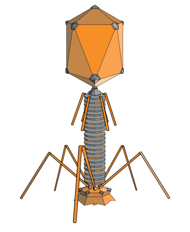
(Figure 1: Common Phage Structure. Figure Source: Wikipedia)

Commonly studied bacteriophages

The classification of phages is complex and numerous. Among countless phages, only a few have been studied in detail. Let’s list a few types of phages to introduce them:

λ phage

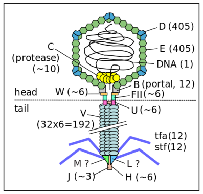
Enterobacteria phage λ (lambda phage, coliphage λ, officially Escherichia virus Lambda) is a bacterial virus, or bacteriophage, that infects the bacterial species Escherichia coli (E. coli).λ Phage is a well-studied temperate bacteriophage that infects the bacterium Escherichia coli (E. coli). Discovered by Esther Lederberg in 1950, it is a key model organism in molecular genetics [2].λ phage has a double-stranded DNA (dsDNA) genome of approximately 48.5 kilobases.λ phage is widely used in molecular cloning and gene regulation studies due to its well-characterized genetics and the ability to integrate into the host genome.

(Figure 2: Structure of λ Phage. Figure Source: Wikipedia)

M13 phage

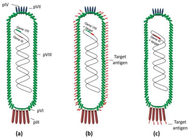
M13 Phage is a filamentous bacteriophage that specifically infects male (F+) strains of E. coli by binding to the F pilus. M13 has a single-stranded DNA (ssDNA) genome of about 6.4 kilobases [3]. M13 is commonly used in phage display technology for protein engineering and antibody development. Its ability to display peptides and proteins on its surface makes it a valuable tool in biotechnological applications.

 ( Figure 3:Scheme of bacteriophage M13: (a) wild-type phage; (b) phage displaying multivalent copies of the target antigen on pVIII protein; and (c) phage displaying multivalent copies of the target antigen on phage pIII protein. In both cases, the DNA fragments encoding the target antigens were cloned into a phage vector [4].)

T4 phage

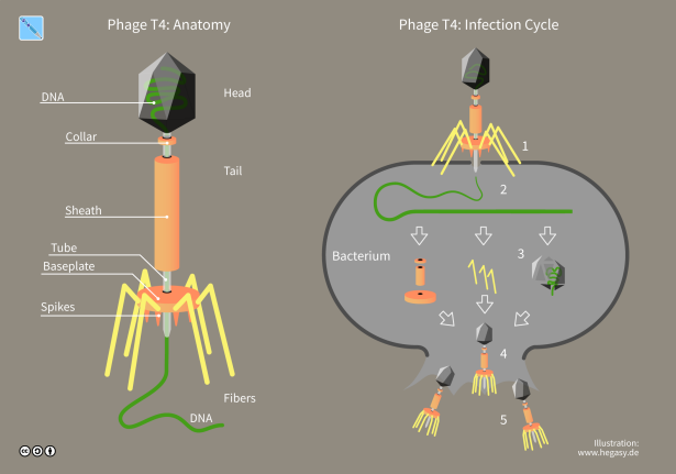
T4 Phage is a lytic bacteriophage that infects E. coli. It is one of the largest and most complex phages studied. T4 has a large dsDNA genome of approximately 169 kilobases. T4 is a relatively large virus, approximately 90 nm wide and 200 nm long (most viruses range from 25 to 200 nm in length) [5]. T4 phage is used as a model system for studying DNA replication, recombination, and repair. Its well-defined life cycle and genetic system make it ideal for these studies.

(Figure 4: Structure of T4 phage. Figure Source: Wikipedia)

T7 phage

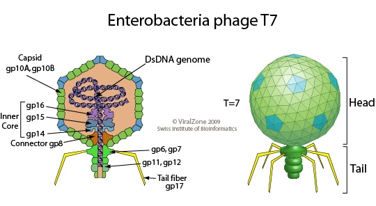
Bacteriophage T7 (or the T7 phage) is a bacteriophage, a virus that infects bacteria. It infects most strains of Escherichia coli and relies on these hosts to propagate. It is known for its simplicity and rapid replication cycle. T7 has a linear dsDNA genome of about 40 kilobases. It is extensively used in molecular biology, particularly in the T7 expression system for high-level protein expression. The T7 RNA polymerase is highly specific and efficient, making it a powerful tool for gene expression studies.

(Figure 5: Structure of T7 phage. Figure Source: Wikipedia)

Phage display technology is an in vitro antibody screening method developed by George P. Smith in 1985. The principle is to insert genes encoding exogenous peptides or proteins into the appropriate positions of the structure genes of the bacteriophage’s outer shell protein. With normal reading frames and without affecting the normal function of the outer shell protein, the exogenous peptides or proteins form fusion proteins on the bacteriophage’s outer shell protein, which is presented on the surface of the bacteriophage as the offspring bacteriophage reassembles. The displayed protein can maintain a relatively independent spatial structure and biological activity, which is conducive to the binding of target proteins. Therefore, target proteins can be quickly used for multi-round screening of phage display antibody libraries and expanded cultivation in E. coli, a process also known as “panning”. Repeated screening can gradually increase the proportion of phages that specifically recognize target molecules, ultimately obtaining peptides or proteins that recognize target molecules.

Reference

Ackermann, H. W. (2009). “Phage Classification and Characterization.” Methods in Molecular Biology, 501, 127-140.

Ptashne, M. (2004). “A Genetic Switch: Phage Lambda Revisited.” Cold Spring Harbor Laboratory Press.

Smith, G. P., & Petrenko, V. A. (1997). “Phage Display.” Chemical Reviews, 97(2), 391-410.

Palma, Marco. (2023). Aspects of Phage-Based Vaccines for Protein and Epitope Immunization. Vaccines. 11. 436. 10.3390/vaccines11020436.

Miller, E. S., et al. (2003). “Bacteriophage T4 Genome.” Microbiology and Molecular Biology Reviews, 67(1), 86-156.

11Jul/24

KMD Bioscience-Deep Analysis of Antibody Discovery Technology-Challenges and Limitations of Hybridoma Technology

The production of monoclonal antibodies using hybridoma technology has advantages such as specificity, consistency, and unrestricted supply, and has a wide range of applications in disease prevention, treatment, and diagnosis. However, the production of monoclonal antibodies using hybridoma technology also has certain limitations. The species for which hybridoma technology is applicable are limited. Traditional hybridoma produces mouse-derived antibodies, which pose certain safety risks when used for human treatment; Secondly, due to the issue of fusion efficiency, it is easy to lose some diversity of B cells, which is not conducive to the discovery of new antibody drugs.

Challenges and Limitations

Immunogenicity

Murine-derived monoclonal antibodies can be recognized as foreign by the human immune system, leading to immune responses against the therapeutic antibodies. This issue has been addressed by creating chimeric, humanized, and fully human antibodies.

Production Complexity

The hybridoma technique involves complex steps that require precise conditions and expertise, making it a time-consuming and costly process.

Ethical Concerns

The use of animals in the immunization step raises ethical concerns. Efforts to develop alternative methods, such as phage display libraries and transgenic animals, are ongoing.

Measures for Improving Restrictions

The field is continually evolving with advancements aimed at improving the efficiency and applicability of monoclonal antibodies. Some of these advancements include:

Antibody Humanization

Techniques to humanize or fully humanize monoclonal antibodies to reduce immunogenicity in patients.

Bispecific Antibodies Production

Development of antibodies that can bind to two targets simultaneously, enhancing therapeutic efficacy.

Gene Editing Technology

CRISPR and other gene-editing tools are being explored to improve the production and functionality of hybridomas.

Recombinant Antibody Technology

Techniques such as phage display, and yeast display are used to produce monoclonal antibodies without the need for hybridoma technology.

Cell-Free Systems

Cell-free protein synthesis systems are being explored to produce antibodies in vitro without the need for living cells, potentially overcoming some of the scaling and ethical issues associated with hybridoma technology.

Hybridoma technology remains a cornerstone in the development of monoclonal antibodies, continually adapting and improving to meet the needs of modern medicine and biotechnology. The continuous advancement of biotechnology aims to address these challenges and provide more efficient and scalable solutions for antibody production.

10Jul/24

KMD Bioscience-Deep Analysis of Antibody Discovery Technology-The Principle and Process of Hybridoma Technology (二)

The entire process of hybridoma has a certain degree of complexity and skill. For example, in the process of immunizing animals, adjuvants are usually used when injecting antigens to avoid non-specific immune reactions that rapidly decompose antigens, achieve slow antigen release and act on the immune system, and ultimately improve the efficiency of producing antibodies with high affinity; For example, as the most crucial step in the preparation of monoclonal antibodies, the cell fusion rate directly affects the preparation of antibodies, especially for the treatment of mouse spleen, which needs to be very careful. A slight negligence can affect the final fusion efficiency.

Hybridoma technology can ensure that the quality of antibodies prepared in different batches is the same and easier to replicate, making it an efficient antibody preparation technique. The technical process for obtaining monoclonal antibodies through hybridoma technology is roughly as follows:

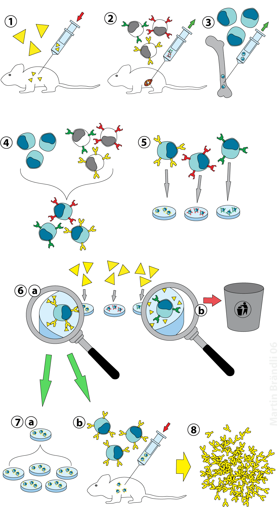
Immunization of Mouse

The process begins with the immunization of a mouse with antigen studied. It stimulates the animal’s immune system to produce a variety of antibodies against the antigen.[1]

Isolation of B cells from the spleen

Spleen cells from the immunized mouse, which contain B lymphocytes producing the antibodies, are harvested.

Cultivation of myeloma cells

Fusion of myeloma and B cells

These cells are then fused with myeloma cells (cancerous B cells) that can grow indefinitely in culture. The fusion is typically facilitated using polyethylene glycol (PEG) or electrical fusion.

Separation of cell lines

The resulting hybrid cells (hybridomas) are cultured in a selective medium, usually hypoxanthine-aminopterin-thymidine (HAT) medium. Only the hybridomas can survive in this medium because they possess the necessary enzyme machinery from the spleen cells to bypass the metabolic block induced by aminopterin.[1]

Screening of suitable cell lines 

Hybridomas are screened for the production of the antibody, typically using assays such as ELISA (enzyme-linked immunosorbent assay). Positive clones are then isolated[1].

In vitro (a) or in vivo (b) multiplication

Obtaining a large amount of monoclonal antibodies through in vitro culture or in vivo induction.

Production and Purification

Once a stable hybridoma producing the desired monoclonal antibody is obtained, it can be cultured in large quantities. The monoclonal antibodies are then harvested from the culture medium and purified using techniques like protein A/G affinity chromatography.

Figure 1: (1) Immunization of a mouse (2) Isolation of B cells from the spleen (3) Cultivation of myeloma cells (4) Fusion of myeloma and B cells (5) Separation of cell lines (6) Screening of suitable cell lines (7) in vitro (a) or in vivo (b) multiplication (8) Harvesting (Figure Source: Wikipedia)


Advantages of Hybridoma Technology

Specificity and Uniformity

Monoclonal antibodies produced by hybridomas are highly specific to a single epitope of the antigen, ensuring uniformity in their action. This specificity is crucial for diagnostic and therapeutic applications.

Unlimited Supply

Hybridoma cells can be cultured indefinitely, providing a consistent and renewable source of monoclonal antibodies.

Customizability

The technology allows for the customization of antibodies to target specific antigens, which is invaluable in research, diagnostics, and treatment of diseases such as cancer and autoimmune disorders.


Applications of Hybridoma Technology

Therapeutics

Monoclonal antibodies are used in the treatment of various diseases, including cancers (e.g., Rituximab for non-Hodgkin lymphoma), autoimmune diseases (e.g., Infliximab for rheumatoid arthritis), and infectious diseases.

Diagnostics

Monoclonal antibodies are essential in diagnostic tests, such as home pregnancy tests and various ELISA-based assays, due to their high specificity.

Research

In research, monoclonal antibodies are used to detect or quantify specific proteins, study cell signaling pathways, and investigate immune responses.

Reference

[1] Parray HA, Shukla S, Samal S, et al. Hybridoma technology a is versatile method for the isolation of monoclonal antibodies, its applicability across species, limitations, advancement, and future perspectives. Int Immunopharmacol. 2020;85:106639. doi:10.1016/j.intimp.2020.106639

09Jul/24

KMD Bioscience-Deep Analysis of Antibody Discovery Technology-Overview of Hybridoma Technology

Antibody screening is a necessary step in the development of cell therapy drugs such as monoclonal antibody drugs, bispecific antibody drugs, ADC drugs, and CAR-T. Screening out high-quality candidate antibodies can greatly increase the likelihood of successful development of these drugs. Antibody drugs are the most important biological drugs besides monoclonal antibodies and recombinant protein drugs and are also a source of new biological therapies, including bispecific antibodies, cell therapies, gene therapies, ADCs, nucleic acid drugs, etc.

At present, common antibody screening methods include hybridoma technology, phage display technology, and single B cell antibody screening technology.

This article will first provide an in-depth analysis of hybridoma technology, and other techniques will be further interpreted in subsequent articles.

In 1975, the birth of monoclonal antibody technology officially ushered in the flourishing development of antibody drugs. Compared to polyclonal antibodies, monoclonal antibodies have a high degree of antigen recognition specificity and protein production uniformity. They can specifically target and bind to various pathogenic agents for disease treatment, and have potential in the field of disease treatment. The preparation method of monoclonal antibodies not only accelerates the development process of life sciences and medicine but also becomes the core technology of the modern biopharmaceutical industry. It has been widely applied in scientific research, diagnosis, antibody drug development, and other manufacturing fields, benefiting humanity in disease treatment and scientific exploration.

So far, hybridoma technology has developed very maturely and is currently the most widely used technology for preparing antibodies. Among the approved therapeutic antibodies, most of the technical routes use mouse-based hybridoma technology.

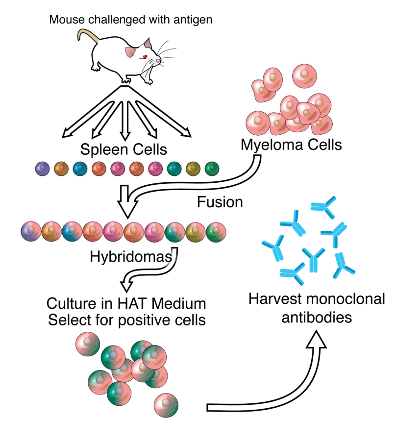
Hybridoma technology is a pivotal technique in biotechnology and immunology for the production of monoclonal antibodies (mAbs). This process first involves injecting antigens that stimulate immune responses into mice (or other mammals). A type of white blood cell, also known as B cell, produces antibodies that bind to the injected antigen. Then, these antibody-producing B cells were collected from the mouse body, and fused with immortalized myeloma cancer cells to produce a hybrid cell line called hybridoma, which has both the antibody-producing ability of B cells and the characteristic of longevity. And the reproductive ability of bone marrow tumors.

Figure 1: A general representation of the hybridoma method used to produce monoclonal antibodies. (Figure Source: Wikipedia)

Hybridoma technology, developed by Georges Köhler and César Milstein in 1975, revolutionized the field by enabling the creation of antibodies that are uniform in structure and specificity. They shared the Nobel Prize of 1984 for Medicine and Physiology with Niels Kaj Jerne, who made other contributions to immunology. The term hybridoma was coined by Leonard Herzenberg during his sabbatical in César Milstein’s laboratory in 1976–1977.[1]

Reference

[1] Milstein, C (1999). “The hybridoma revolution: an offshoot of basic research”. BioEssays. 21 (11): 966–73.


08Jul/24

Antibody Drugs and Antibody Discovery-KMD Bioscience

Overview of Antibody Drugs

Compared to traditional small molecule drugs, antibody drugs offer several distinct advantages. Firstly, antibody drugs can target a broader range of biological molecules, including proteins, cell surface receptors, and antigens. This expanded range of targets allows for more precise intervention in various diseases, such as cancers, autoimmune disorders, and infectious diseases. Additionally, antibody drugs are less likely to induce drug resistance. This is largely because antibodies bind specifically and with high affinity to their targets, which reduces the likelihood of mutations that confer resistance.

Moreover, the affinity of antibody drugs can be engineered to enhance their effectiveness and safety. By improving the binding affinity of antibodies to their targets, therapeutic efficacy can be increased while minimizing off-target effects, which in turn reduces toxicity and side effects. This engineering can involve modifications to the antibody structure to increase its stability, prolong its half-life in the bloodstream, and improve its interaction with the immune system.

In summary, the versatility, specificity, and engineering potential of antibody drugs makes them a powerful tool in modern medicine, providing more treatment options with potentially fewer side effects and a lower risk of resistance compared to traditional small molecule drugs.

Statistics on FDA-Approved Antibody Drugs

As of 2024, the FDA has approved over 100 antibody drugs in total. This milestone was achieved in 2021 when the FDA approved the 100th monoclonal antibody drug dostarlimab [1]. This growth shows the importance and popularity of antibody drugs in the treatment field, especially in the treatment of cancer, immune diseases, and infectious diseases.

In 2023 and early 2024, the FDA continued to approve multiple new antibody drugs, including lecanemab (Leqembi) for the treatment of Alzheimer’s disease, elanatamab (Elrexfio) for multiple myeloma, and rosanolixizumab (Rystiggo) for generalized myasthenia gravis. [2]

Table 1. Commercially sponsored monoclonal antibody therapeutics granted a first approval in any country during 2023.[3]

INN (Brand Name) Target; Format Indication First Approved
Lecanemab (Leqembi) Amyloid beta protofibrils; Humanized IgG1ҡ Early Alzheimer’s disease
Rozanolixizumab (RYSTIGGO) FcRn; Humanized IgG4ҡ Generalized myasthenia gravis
Pozelimab (VEOPOZ) Complement C5; Human IgG4ҡ CHAPLE disease
Mirikizumab (Omvoh) IL-23p19; Humanized IgG4ҡ Ulcerative colitis
Talquetamab (Talvey) GPCR5D, CD3; Humanized IgG4ҡ Multiple myeloma
Elranatamab (Elrexfio) BCMA, CD3; Humanized IgG2ҡ bispecific Multiple myeloma
Epcoritamab (EPKINLY) CD20, CD3; Humanized IgG1ҡ/λ bispecific Diffuse large B-cell lymphoma
Glofitamab (COLUMVI) CD20, CD3e; IgG1ҡ/λ bispecific Diffuse large B-cell lymphoma
Retifanlimab (Zynyz) PD-1; Humanized IgG4ҡ Merkel cell carcinoma
Concizumab (Alhemo) Tissue factor pathway inhibitor; Humanized IgG4ҡ Hemophilia A or B with inhibitors
Lebrikizumab (EBGLYSS) IL-13; Humanized IgG4ҡ Atopic dermatitis
Tafolecimab (SINTBILO) PCSK9; Human IgG2ҡ Primary hypercholesterolemia and mixed dyslipidemia
Narlumosbart (Jinlitai) RANKL; Human IgG4ҡ Giant cell tumor of bone
Zuberitamab (Enrexib) CD20; Chimeric IgG1ҡ Diffuse large B-cell lymphoma
Adebrelimab (Arelili) PD-L1; Humanized IgG4ҡ Extensive-stage small cell lung cancer
Divozilimab (Ivlizi) CD20; Humanized IgG1ҡ Multiple sclerosis


The discovery of antibodies, including phage display antibody screening or yeast screening, antibody sequencing, and subsequent antibody validation evaluation. Among them, antibody screening, as a crucial step, takes the longest time and has low screening efficiency, which also limits the cycle of antibody discovery. In recent years, the rapid development of antibody screening technology has greatly shortened the entire process of antibody discovery.

KMD Bioscience has a professional antibody discovery platform, where customers can choose appropriate antibody screening techniques according to their different needs. Our professional scientists can provide customized solutions, and in subsequent articles, we will deeply analyze antibody discovery techniques. You can follow KMD Bioscience’s official website for more information.

Reference

https://www.antibodysociety.org/antibody-therapeutics-product-data/

Crescioli, S., Kaplon, H., Chenoweth, A., Wang, L., Visweswaraiah, J., & Reichert, J. M. (2024). Antibodies to watch in 2024. mAbs, 16(1). https://doi.org/10.1080/19420862.2023.2297450

05Jul/24

KMD Biosciecne-De novo sequencing principle of protein

De novo sequencing refers to the process of determining the amino acid sequence of a protein from scratch, without any prior knowledge of its DNA sequence. This technique is crucial for studying proteins from organisms with unsequenced genomes or proteins with post-translational modifications. Here’s an outline of the experimental principle behind protein de novo sequencing:

Step 1 Protein Isolation and Purification

The first step involves isolating the target protein from a complex mixture and purifying it to a sufficient degree to allow for accurate sequencing.

Step 2 Protein Reduction and Alkylation (optional)

If the protein contains disulfide bonds, they may be reduced to free thiol groups and then alkylated to prevent the re-formation of the disulfide bonds. This step linearizes the protein structure and makes it more amenable to enzymatic or chemical cleavage.

Step 3 Protein Digestion

The purified protein is enzymatically or chemically digested into smaller peptide fragments. Common proteolytic enzymes used for digestion include trypsin and chymotrypsin, which cleave proteins at specific amino acid residues. This digestion results in a mixture of peptides with known cleavage sites.

Step 4 Peptide Separation

The resulting peptide mixture is often separated using chromatographic techniques such as liquid chromatography (LC) to reduce complexity and enable more accurate sequencing.

Step 5 Mass Spectrometry Analysis

The separated peptides are analyzed by mass spectrometry (MS). The mass spectrometer measures the mass-to-charge ratio of the peptides and their fragments, generating a spectrum that reflects the peptide’s composition and sequence.

Step 6 Peptide Fragmentation

Within the mass spectrometer, peptides are fragmented along the backbone, generating a series of ionized fragments. This process is often achieved through techniques like Collision-Induced Dissociation (CID) or Electron Transfer Dissociation (ETD).

Step 7 Spectrum Analysis

The generated spectra are analyzed to determine the sequence of amino acids in each peptide. The analysis involves matching the observed fragment ion masses to theoretical fragment ion masses derived from all possible amino acid sequences.

Step 8 Sequence Assembly

The amino acid sequences of individual peptides are assembled to deduce the complete sequence of the original protein. Overlapping regions between peptides help in accurately assembling the full sequence.

Step 9 Database Searching and Sequence Alignment (if applicable)

While de novo sequencing is intended for proteins with unknown sequences, any obtained sequence information can be compared with known sequences in databases to find homologies or to confirm the de novo sequencing results.

Step 10 Validation

Additional experiments or analyses may be conducted to validate the obtained protein sequence, such as synthesis and characterization of the protein based on the deduced sequence or comparison with similar known proteins.

De novo sequencing is a complex but powerful technique that enables the exploration and characterization of proteins in the absence of genomic information, thus playing a crucial role in proteomics and related fields of research.

KMD Bioscience has been dedicated to protein expression and sequencing research for over 10 years. Based on high-resolution mass spectrometry, We can provide customers with high-fidelity antibody and protein sequencing services. KMD Bioscience has highly skilled laboratory technicians with extensive experience in protein and antibody sequencing and can provide mass spectrometry-based protein sequencing analysis services. Combined with bioinformatics databases, our sequencing platform can realize rapid and accurate analysis of protein primary structure, and can also provide protein N-terminal sequencing services. For more information, Visit us at  https://www.kmdbioscience.com/ to have a detailed understanding.

04Jul/24

KMD Bioscience-Immunoprecipitation: Applications and Protocols

Immunoprecipitation (IP) is a widely employed technique in molecular biology and biochemistry, enabling the isolation and concentration of specific proteins from complex mixtures. By leveraging the specificity of antibodies to selectively bind proteins, IP facilitates the study of protein-protein interactions, protein modifications, and protein functions. This article delves into the myriad applications of immunoprecipitation and outlines some common protocols utilized in this technique.

The essence of immunoprecipitation lies in the interaction between the antigen (target protein) and the antibody. A suitable antibody is chosen to selectively bind the protein of interest, forming an antigen-antibody complex. This complex is then precipitated from the solution, typically using protein A/G agarose beads. The beads are collected, and washed to remove unbound materials, and the bound proteins are eluted for further analysis. Understanding the core principles and methodology of IP is pivotal for obtaining reliable and interpretable results.

Immunoprecipitation (IP) and other protein interaction applications such as yeast two-hybrid (Y2H), protein microarrays, and Förster Resonance Energy Transfer (FRET) serve the common goal of elucidating protein interactions, albeit through distinct mechanisms. IP leverages the specificity of antibodies to isolate and concentrate particular proteins from a mixture, providing a direct method of assessing protein interactions or modifications in a near-native state. On the other hand, Y2H employs a genetic system in yeast to detect interactions, offering a high-throughput albeit less direct method, which may miss interactions occurring outside the yeast’s nuclear environment. Protein microarrays provide a high-throughput platform to study protein-protein interactions on a large scale, yet may not always reflect the natural cellular context. FRET utilizes energy transfer between two light-sensitive molecules to gauge the interaction and distance between proteins, offering real-time interaction analysis within living cells. Each of these methods has its own set of advantages and limitations concerning sensitivity, throughput, and contextual relevance, choosing method contingent on the specific requirements of the research question at hand.

Immunoprecipitation finds extensive applications in research fields like oncology, immunology, and neurobiology. It is instrumental in studying protein-protein interactions, identifying post-translational modifications, and investigating cellular signaling pathways. Moreover, IP serves as a crucial step in various downstream applications such as Western blotting, mass spectrometry, and enzyme activity assays which provide insights into the functional and structural aspects of proteins.

Here’s a brief protocol of Immunoprecipitation:

Step 1 Sample Preparation

Preparing a clear, homogenous sample is the first step, which involves lysing cells or tissues to release proteins. Adding protease and phosphatase inhibitors to prevent protein degradation.

Step 2 Antibody Selection and Binding

Choosing a high-affinity, specific antibody is crucial. The antibody is incubated with the sample to allow binding to the target protein. Incubate at an appropriate temperature for a specified time (e.g., 1-2 hours) to allow antibody-protein binding.

Step 3: Immobilization to a Solid Support

The antibody-protein complexes are then immobilized on a solid support, usually beads coated with protein A or G which bind the Fc region of antibodies. During this incubation, the antibody binds to its target protein, and the protein-bead-antibody complex forms.

Step 4 Washing and Elution

Washing the beads several times with the wash buffer to remove unbound proteins and contaminants, and the target protein is eluted from the beads, often by altering the pH or using a competitive ligand.

Step 5 Analysis

The eluted protein can be analyzed using various techniques like Western blotting, mass spectrometry, or gel electrophoresis.

Step 6 Optimization

Fine-tuning parameters such as incubation times, temperatures, and buffer compositions is often necessary to achieve optimal results.

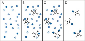
Figure 1Immunoprecipitation. (A) Before an immunoprecipitation procedure begins, a protein of interest exists in a heterogenous sample with many other proteins. (B) A scientist adds antibodies that recognize the protein to small beads and then adds the beads to the sample. (C) The antibodies bind to the proteins, causing them to precipitate out of solution and adhere to the beads. (D) The beads are removed from the sample, purifying the protein of interest. The proteins can be removed from the beads by changing the salinity or pH of the solution.(Figure source: Matt Carter, Jennifer C. Shieh, 2010)


Immunoprecipitation remains a cornerstone technique, bridging the molecular interactions with the vast expanse of research insights. The meticulous adherence to protocols coupled with a profound understanding of its applications propels the scientific inquiry forward, opening avenues for novel discoveries and a deeper understanding of life’s molecular machinery.


KMD Bioscience has been dedicated to the study of protein-nucleic acid interactions for over 10 years. Eukaryotic genomic DNA exists in the form of chromatin, and the study of protein-DNA interactions in the chromatin environment is a fundamental way to elucidate the mechanisms of eukaryotic gene expression. Scientists in KMD Bioscience, which have accumulated a wealth of experience in immunoassay technology, are able to quickly customize protein-nucleic acid interaction study assays for different clients. With years of technical expertise, KMD Bioscience is not only able to complete the experimental content quickly, but also to ensure that each step of the experiment is carefully controlled to ensure that the test results are accurate, objective and credible. KMD Bioscience, which has complete protein detection equipment, has established mature and perfect antibody platforms, protein platforms and other technology platforms.

For more information, Visit us at  https://www.kmdbioscience.com/ to have a detailed understanding.