0512 月/23

Production process of nanobodies (二): Antibody screening and identification process

The previous article talked about the first stage of nanobody production, and this article mainly talks about the latter part of antibody production. The specific procedure is as follows:

Coated immune tube:

50ug antigen was added to 2mL PBS and added to the immune tube for overnight incubation at 4℃.

Isolation:

The amplified and purified phage was added to 1mL 3% BSA and incubated for 2h at room temperature. At the same time, 2-3mL 3% BSA was added to the coated immune tubes and incubated at room temperature for 2h.

Antigen and bacteriophage incubation:

The sealed immune tube was washed 3 times with PBS containing 0.01% Tween for 5 minutes each time. The closed phage library was added to the closed immune tube, PBS was added until 2-3mL, and the phage library was incubated at room temperature for 1h.

Cleaning:

After incubating the antigen and phage, the immune tube containing 0.1% Tween was washed 20 times with PBS for 5 minutes each time.

Elution:

1mL 100mM Trimethymime was added to the immune tube, incubated at room temperature for 10 minutes, 1M Tris-HCl was added to neutralize Trimethymime, and the last 1.5mL eluted phage was transferred to a new centrifuge tube. The eluted phage was amplified and purified according to the phage library, and then the screening process was repeated twice after amplification, and the amount of antigen covered by the immune tube was halved successively to obtain the eluted phage after 3 screening. The elution phage can be sequenced by NGS to obtain the candidate nanoantibody DNA sequence library.

ELISA identification:

At the same time, after gradient dilution of the bacteriophages obtained in the previous step, 100ul of each bacteriophage was added to TG1 bacterial solution with OD600nm of 0.5, cultured at 37℃ for 30 minutes, coated with 2x YT culture plate containing ambenomycin, and incubated at 37℃ overnight for the next day to obtain monoclonal colonies. At least 192 single colonies were randomly selected onto 96-well cell culture plates containing 2x YT culture solution and cultured overnight at 37℃ as seed bacteria plates, and the bacterial solution 2ul was re-added to the new 96-well plates (each well containing 200ul 2x YT fresh culture solution). 100ug/ml ammobenzyl) 5 hours later, the auxiliary phage was added into the culture hole, and the final concentration of 50ug/ml kanamycin was added at 37℃ for 30 minutes, and the culture was incubated overnight at 30℃. On the second day, the bacteria solution after overnight culture was centrifuged to obtain the supernatant containing phage.

The overnight coated antigen hole and the BSA coated control hole were sealed with 3% BSA, then the bacteriophage supernatant obtained in the previous step was added, and incubated at room temperature for 1h. After three times of cleaning with PBS containing 0.1% Twain, the phage antibody was added for incubation, and the light absorption value of each well was read at a wavelength of 450nm after color development with TMB. Colonies with large absorbency ratio between antigen-coated pores and corresponding control pores were selected for sequencing (two phage-Elisa analyses independently) to obtain the gene sequence of the nanobodies.

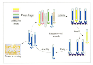
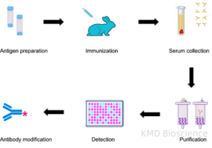
Fig. 1 Antibody screening and identification process

KMD Bioscience provides customized screening system for customers. According to different needs of customers, we provide solid phase screening, solid phase/liquid phase screening and cell screening methods. KMD Bioscience has established a complete and mature phage antibody display technology platform.In addition, KMD Bioscience has rich experience in antibody engineering construction, and can provide three-dimensional antibody upstream and downstream services, including antibody humanization service, human scFv antibody library construction service, human Fab antibody library construction service, human antibody phage library Production service, phosphorylated antibody customization service, antibody affinity maturation service, etc.

2811 月/23

Production process of nanobodies (一): Alpaca immune and Phage construction process

From the brief overview of Nanobody production in the previous article, we understand the general process. Next, we will spend two articles introducing the specific production process. First, we will introduce the previous part of Nanobody production from alpaca immunization to library construction.

Alpaca immune process

Antigen Production:

an alpaca can be immunized with 1-3 antigens at the same time, the total amount of antigen for each immunization is maintained between 1-2mg, the volume is below 2mL, and the antigen and adjuvant are 1:1 emulsified before immunization to form a uniform mixture, which is stored at 4℃.

Immunizing alpacas:

After recording a blank alpaca ear number, the immunization experiment was started. Each time, the alpaca was injected into the left and right sides near the neck lymph nodes, and each side was injected into 2 points, and about 0.4mL of emulsified antigen was injected into each point. Observation for half an hour after immunization confirmed that the alpaca was in good condition and had no discomfort symptoms. Immunizations are given every 2 weeks, at least 4 times.

Blood collection:

50ml of blood was collected from the neck vein of alpaca at an interval of 5-7 days after the fourth immunization.

Serum separation:

Before each antigen immunization, blood was taken for immune evaluation, 5mL of blood was taken each time; On the same day, the blood was precooled by a 25℃ centrifuge and centrifuged at 400 xg for 30 minutes to separate and preserve the upper serum for subsequent antibody titer detection.

Isolation of lymphocytes:

15mL of cell separation solution was first added to a 50mL centrifuge tube, and then 15mL of blood was slowly added. Add blood slowly and carefully to prevent mixing of blood and separation solution. Then the centrifuge was precooled to 25℃ and centrifuged at 400 xg for 30 minutes. The separation of blood in the centrifuge tube was observed. The upper level serum was stored in the new centrifuge tube at -80 ℃. Each tube was added with 10mL PBS buffer at room temperature and centrifuged at 400 xg at 25℃ for 20 min. The supernatant was removed, 5mL of PBS buffer at room temperature was added to each tube, and the number of cells was calculated using a blood cell counting plate, and then centrifuged at 25℃ and 400 xg for 20 min. The supernatant was removed, and the isolated lymphocytes were dissolved with RNAiso Plus according to the number of cells to obtain 107/mL cytolytic solution, which was stored at -80℃.

Phage construction process

RNA extraction:

Peripheral blood lymphocytes preserved with Trizol were dissolved on ice and transferred to a 1.5mL centrifuge tube, where 1/5 volume of chloroform was added to shake and mix. Stand at room temperature for 5 minutes at 4℃, centrifuge at 12000g for 15 minutes; Transfer the supernatant after centrifugation to a new centrifuge tube; Isopropyl alcohol of equal volume was added to the new centrifuge tube. Let stand at room temperature for 10 minutes and centrifuge at 4℃ 12000g for 10 minutes; Clean and precipitate with 75% ethanol, centrifuge at 4℃ 7500g for 5 minutes, discard the supernatant, precipitate and dry at room temperature, and dissolve in appropriate amount of RNA-free water.

Reverse transcription cDNA:

According to the instructions of the reverse transcription kit, the RNA obtained in the previous step was divided into two parts and reverse-transcribed into cDNA. The reverse transcription primers were Oligo T and random primers, respectively.

Amplification of antibody fragments:

Specific antibody fragments are amplified from reverse-transcribed cDNA, and PCR amplification is performed using Taq DNA Polymerase Hot Start enzyme.

Two rounds of PCR reaction were performed, and the obtained PCR amplification products were recovered using DNA purification recycling kit according to the instructions.

Cloning to phage plasmid:

The diversified antibody gene sequence and phage vector amplified in the previous step are enzymically cut, respectively purified and linked. The connected products were recovered by DNA purification recycling kit according to the instructions, and dissolved in ultra-pure water.

TG1 conversion:

Place the cup on ice for pre-cooling, wait for 100ul of TG1 receptive cells to melt and add 100ng of the recovered connection products, transfer the mixed receptive cells and connection products to the pre-cooled cup, use the Bacteria transformation procedure preset by the tester for electric shock transformation, immediately add 1mL SOC culture medium into the cup. After at least 20 revolutions, the cells were resuscitated at 37℃ for 60 minutes and coated on a LB culture plate containing ampicillin resistance for overnight growth. The cells on the culture plate after overnight growth in the previous step were washed and scraped off with 2xYT medium and a coating rod, and then stored at -80℃ after adding 20% glycerin.

Amplification and purification of phage library:

After mixing the bacteria scraped from the previous step, about 10^9 bacteria were transferred to 100mL 2x YT culture medium pre-added with ampicillin antibiotics, and cultured at 220rpm at 37℃ until OD600nm reached 0.5. According to the ratio of helper phage: the number of bacterial cells was 20:1, the auxiliary phage was added and continued to be cultured at 37℃ for 30 minutes. The final concentration of kanamycin was 50ug/ml and cultured in shaking bed at 30℃ overnight. The bacteria cultured overnight were centrifuged at 13000rpm at 4℃ for 5 minutes, the supernatant was transferred to a new centrifuge tube, and 1/4 volume of pre-cooled 5x PEG8000/NaCl was added, and incubated on ice for 30 minutes. Centrifuge at 4℃ at 13000rpm for 10 min to remove the supernatant and add 1mL PBS buffer to dissolve the precipitation. 250ul 5X PEG8000/NaCl was added again and incubated on ice for 10 minutes. After centrifugation at 4℃ at 16000g for 15 minutes, the supernatant was removed and the precipitation was dissolved in 1mL PBS to obtain the phage library.

In the next article, we will talk about the second half of nanobody production, which is library screening and antibody identification.

Fig.1 Alpaca immune and Phage construction process

KMD Bioscience provides healthy adult alpacas (28-36 months old) for immunization, and high-quality libraries and monoclonal antibody cell lines can be obtained in about 16-20 weeks, and complete experimental process records and immune dynamic videos can be delivered. According to the antigen type of customers, KMD Bioscience will make specific immune antigen generation plans and immunization plans. KMD Bioscience displayed VHH antibody (Nanobody) with pIII protein of M13 phage, prepared TG1 transduced competent cells, constructed phage vector, and measured phage storage capacity. (Learn more about antibody discovery services)

This article serves as a reference material for enthusiasts in scientific research. It does not substitute for professional knowledge or hands-on experimental procedures which require more detailed and professional information. In case of any content infringement, kindly reach out to the author for immediate deletion of the contentious material.

Reference

Muyldermans S. Nanobodies: natural single-domain antibodies. Annu Rev Biochem. 2013;82:775-97.

2011 月/23

Antibody Generation Techniques: Polyclonal Antibody Production

Antibodies, as pivotal components of the immune system, have been employed in various scientific and medical fields. The generation of antibodies, as a critical aspect of immunological responses, has garnered significant attention in biomedical research and clinical applications.

Polyclonal Antibody Production procedure

Polyclonal antibodies (pAbs) are a diverse mixture of antibodies produced by different B cell lineages within an immunized host, typically a rabbit or goat. The process begins with the administration of the target antigen, following which the host’s immune system generates a broad spectrum of antibodies recognizing multiple epitopes on the antigen. The antiserum, containing this heterogeneous antibody population, is then collected and may undergo further purification to obtain the desired antibody fraction.

The general procedure to produce polyclonal antibodies is as follows:

1 Antigen preparation

2 Adjuvant selection and preparation

3 Animal selection

4 Injection and ELISA titer testing

5 Blood serum extraction and antibody purification.

Fig1. Polyclonal Antibody Production Procedure

Diagnostic Applications of Polyclonal Antibodies (pAbs)

——Broad Reactivity: pAbs can recognize multiple epitopes on an antigen, which is beneficial in diagnostics where a broader recognition spectrum is advantageous.

——Use in ELISA: Like mAbs, pAbs are also utilized in ELISA and other diagnostic assays, especially when detecting a range of antigenic variants is crucial.

Therapeutic Applications of Polyclonal Antibodies

——Antivenom Production: pAbs play a crucial role in antivenom production, where a broad spectrum of antibodies is required to neutralize various toxins present in venom.

——Infectious Diseases: They are also utilized in passive immunotherapy for infectious diseases, providing a broad-spectrum immune response.

KMD Bioscience‘s scientists are experts working for years in antibody production and thanks to their experience and expertise, a mature antibody production platform with advanced has been built. With the support of our reliable antibody platform, KMD Bioscience can provide a full range of services for your polyclonal antibody research, covering antigen design, animal immunization, serum collection, titer analysis, and final antibody purification, every step of polyclonal antibody development.


2011 月/23

Protein Production Process-Recombinant protein expression

1. What is recombinant protein expression?

It reveals that life activities can be explored from different levels such as genes and proteins. With the continuous development of science and technology, protein research has developed from protein extraction to recombinant protein expression. At present, we can use a variety of recombinant protein expression systems (prokaryotic expression system, yeast expression system, insect expression system, mammalian expression system, plant expression system) to recombinant expression, separation and purification of our target proteins to further explore the function of proteins, providing a convenient method for scientific research (Belenkaya S. V, 2023).

2. Experimental procedure of recombinant protein expression

The main components include expression vectors and host cells, and the main experimental procedures are as follows:

(1) Construction of expression vector.

The selection of expression vector is usually based on the purpose of the expressed protein, known information and cloning and purification strategies. After the vector is selected, the target gene is connected to the expression vector by homologous recombination or seamless cloning.

(2) Transform host cells.

Host cell selection can be based on the characteristics of the host strain as well as the use of the target gene expression product. For example, when there are more proteases produced in the host cell, which affects the stability of the target gene expression product, the protease-deficient strain can be selected, such as BL21 series is suitable for the expression of toxic proteins. When the foreign gene to be expressed is a eukaryotic gene, which is difficult to be expressed in prokaryotes, the Rosetta series strains supplemented with rare codons can be selected. After selecting a suitable host strain, the constructed plasmid containing the target gene is transformed into the host cell, and the positive clone is screened.

(3) Induced expression of target protein

Culture conditions are the key factors for successful production of recombinant proteins. IPTG is the commonly used induction method for prokaryotic expression, and methanol is the commonly used induction method for yeast expression. However, exploration and optimization should be carried out for different target genes and host strains, the concentration of inducers, and the culture temperature and time to achieve the best culture conditions.

(4) Separation and purification of target protein.

According to the properties of proteins, different expression forms and fusion labels, suitable methods were selected to isolate and purify the target proteins.

(5) Detection and identification of target protein

The size and purity of purified protein can be detected by SDS-PAGE and Western-blolt methods. All procedures should be performed on ice to prevent protein denaturation.

KMD Bioscience has been committed to the expression, separation and purification of recombinant proteins for many years, and has built protein platforms such as prokaryotic expression system, yeast expression system, insect expression system, mammalian expression system and plant expression system for different protein expression needs. For protein expression purification, we can provide the following services and protein expression solutions.

This article serves as a reference material for enthusiasts in scientific research. It does not substitute for professional knowledge or hands on experimental procedures which require more detailed and professional information. In case of any content infringement, kindly reach out to the author for immediate deletion of the contentious material.

References:

Belenkaya S. V., et al.”Comparison of the Biochemical Properties of Recombinant Alpaca (Vicugna pacos) Chymosins Produced in Pro- and Eukaryotic Expression Systems.”Applied Biochemistry and Microbiology 59.5(2023):630-635.

Suo Qian, et al.”[Prokaryotic expression, polyclonal antibody preparation, spatio-temporal expression profile and functional analysis of c-Myc of Helicoverpa armigera (Lepidoptera: Noctuidae)]..” Sheng wu gong cheng xue bao = Chinese journal of biotechnology39.7(2023):2730-2742.

Wu Yijian, et al.”An outlook to sophisticated technologies and novel developments for metabolic regulation in the Saccharomyces cerevisiae expression system.”Frontiers in Bioengineering and Biotechnology 11.(2023):1249841-1249841.

2011 月/23

Overview: How Nanobody is Produced

Compared with traditional monoclonal antibodies, the Production of nanobody has great advantages. It can be summarized as immunizing alpacas to obtain antibody genes and phage display to obtain target antibody sequences. It is mainly divided into four steps: alpaca immunization, phage library construction, antibody library screening, antibody expression and functional verification.

In order to build an immune pool, young, healthy llamas, dromedaries, alpacas, etc. must first be immunized. Typically, animals are injected 4-8 times with a mixture of target antigens and adjuvants over a 2-month time span. Generally, each injection is about 50-200μg immunogen; The exact amount obviously depends on the molecular weight of the antigen, but more on its immunogenicity and/or toxicity.

Immunogen type: The use of soluble, correctly folded recombinant proteins is preferred, and there is also direct DNA immunity. Up to 10 proteins are usually mixed to immunize animals, but more complex mixtures have also been used (e.g., protein extracts from viruses, bacteria, parasites, whole mouse spleen cells, or cancer cells).

To increase the likelihood of a specific epitope successfully acquiring a nanobodies, immunizing more than one animal is recommended. They are inbred animals, and each one produces a unique immune response, obtaining a larger panel of nano antibodies from which to select the best-performing nanobodies.

Fig. 1 Brief procedure of nanobody production

Some studies have shown a higher proportion of HCAb antibodies than classical antibodies in camels or dromedaries compared to llamas and alpacas. After immunization, 50-100 ml of anticoagulant blood (usually from the jugular vein, although lymph node biopsy is also a good raw material) is taken, lymphocytes are prepared and mRNA is extracted. The mRNA is converted to cDNA for amplification of the VHH gene (two-step nested PCR).

Some molecules, such as RNA or DNA, are not immunogenic and cannot stimulate the production of HCAb, or some compounds are too toxic, infectious, or harmful to animals or the environment, in which case a natural library of nano antibodies or a synthetic library of nano antibodies can be envisioned (Erwin De Genst, 2006).

In order to be able to screen high-affinity nano antibodies, the size of the natural antibody library is generally 109-1010, of which about 80% should encode nano antibodies. To build such a large and diverse library of natural nano antibodies, blood needs to be collected from multiple animals (at least 10 animals to avoid bias in HCAbs due to allergies or previous infections). There are an estimated 106 lymphocytes per mL of blood, and given that only a small fraction of them are B cells, about 50% of which may express HCAb, 10L of blood is needed to build a nanoantibody library of 1010 different VHH clones.

Combining in vitro antibody maturation techniques, such as DNA recombination, error-prone PCR, and random primers, can increase the diversity of complementary determining regions (CDRs), as well as the specificity and affinity of the nanobodies. Phage display and ribosome display are two main strategies for screening nanomes from natural antibody libraries (Rossotti MA,2023).

KMD Bioscience has established a complete and mature phage antibody display technology platform. In addition, KMD Bioscience has rich experience in antibody engineering construction, and can provide three-dimensional antibody upstream and downstream services, including antibody humanization service, human scFv antibody library construction service, human Fab antibody library construction service, human antibody phage library Production service, phosphorylated antibody customization service, antibody affinity maturation service, etc.(Learn more about antibody discovery services)

This article serves as a reference material for enthusiasts in scientific research. It does not substitute for professional knowledge or hands-on experimental procedures which require more detailed and professional information. In case of any content infringement, kindly reach out to the author for immediate deletion of the contentious material.

Reference

De Genst E, Saerens D, Muyldermans S, Conrath K. Antibody repertoire development in camelids. Dev Comp Immunol. 2006;30(1-2):187-98.

Rossotti MA, Trempe F, van Faassen H, Hussack G, Arbabi-Ghahroudi M. Isolation and Characterization of Single-Domain Antibodies from Immune Phage Display Libraries. Methods Mol Biol. 2023;2702:107-147.

1511 月/23

Nanobodies: A New Era in Drug Discovery and Development

1. What is Nanobody?

Nanobody was first proposed and reported in Nature by Belgian scientists in 1993. The traditional antibody molecule (IgG) is composed of two identical heavy chains and light chains. The light chain of antibody molecule contains one VL region and one CL region, while the heavy chain has one VH region and three CH regions (CH1, CH2 and CH3).

Alpaca only has heavy chain antibody but lacks light chain, which is composed of four HC’s and two VH’s. Among them, VHH region is also called nanobodies or single domain antibody (sdAb) and Nanobody (Nb). It’s crystal is 2.5nm, 4nm in length and only 15KDa in molecular weight. Nano-antibodies are mainly used in biomedical research and development (genetic engineering drug research and development, ADC drug research and development); Used for clinical diagnosis in vitro (colloidal gold method, enzyme-linked immunosorbent assay, electrochemiluminescence method); It is used in basic research fields such as tumor research and immunology. At present, the development of CAR-T therapeutic antibody has attracted more and more researchers’ attention.

Fig. 1. The structure of Nanobody

2. Advantages of Nanobody

(1) stability at high temperature and pH;

(2)VHH can recognize antigen sites that are not usually recognized by conventional antibodies;

(3) Their small molecular fragments are helpful for rapid tissue penetration and labeling applications, including crossing the blood-brain barrier;

(4) Cost-saving substitutes for mass production.

3. Application of Nanobody (VHH antibody)

Based on the special structure and properties of Nanobody, it is favored by people in the field of CAR-T therapeutic antibody development.

CAR-T therapy is a new type of targeted therapy for tumor, that is, chimeric antigen receptor T cell immunotherapy. By means of genetic engineering technology, activated T cells are equipped with a positioning navigation device CAR (tumor chimeric antigen receptor) to turn T cells into “super soldiers”, that is, CAR-T cells. With its positioning navigation device CAR, tumor cells in vivo can be accurately and specifically identified and eliminated.

KMD Bioscience can provide a variety of camel-derived Nanobody development services, including alpacas, llamas, camelus, etc. Based on the phage display technology platform, KMD Bioscience can provide major experimental links including antigen design, alpaca immunity, library construction and screening, and activity function identification, and provide alpaca VHH antibodies with high specificity and high affinity for scientists around the world.

This article serves as a reference material for enthusiasts in scientific research. It does not substitute for professional knowledge or hands-on experimental procedures which require more detailed and professional information. In case of any content infringement, kindly reach out to the author for immediate deletion of the contentious material.