186 月/24
Antibody Discovery-KMDBioscience

Alpaca immune isolation PBMC common problems and solutions-KMD Bioscience

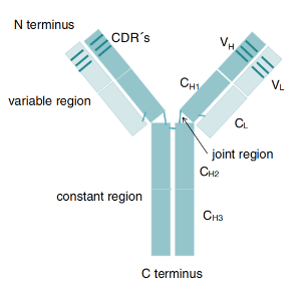
Alpaca Immune isolation What cells are isolated by PBMC?

The isolated peripheral blood mononuclear cells are the main cell types of mononuclear cells in the blood, mainly including lymphocytes (T /B), monocytes, phagocytes, dendritic cells, and other small numbers of cell types, of which lymphocytes account for a large part.

What is the purpose of PBMC separation?

The main purpose of isolating PBMC is to remove multinucleated cells and red blood cells so that it can easily simulate the blood immune environment in vitro. Preparation of PBMC from blood is a common step before isolating a specific subpopulation of immune cells.

What are the precautions during PBMC separation?

(1) Not applicable to abnormal samples such as blood clotting or samples over 48 hours.

(2) It is recommended to use 4-9ml samples for 15ml separation tubes; For 50ml separation tubes, 13-30ml samples are recommended.

(3) For samples placed for more than 24 hours, it is recommended to lengthen the centrifugation time.

(4) Before harvesting cells after centrifugation, the upper plasma layer can also be absorbed, collected, or discarded to help prevent platelet contamination.

(5) When pouring supernatant after centrifugation, do not invert the separation tube for more than 2s.

(6) Do not reuse the separation pipe.

(7) After centrifugation, cells may gather on the wall of the separation tube above the enrichment layer. This polymerization is normal and is affected by sample quality, sample placement time, and type of anticoagulant. This polymerization is independent of the use of the separation tube. The cells can be removed by lightly scraping one side of the cluster using the pipette tip.

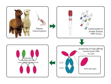
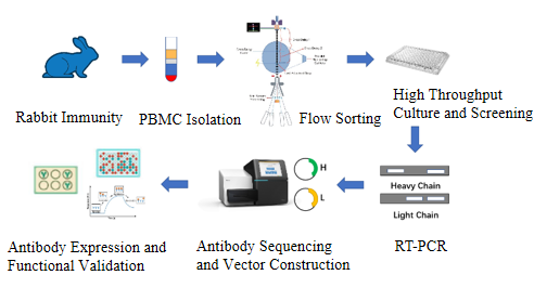
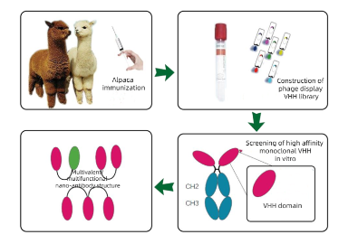
How can alpaca Immunology obtain highly effective serum and high-affinity antibodies through follow-up screening?

High-quality immunogen preparation

the purity of the immunogen should be as high as possible to avoid non-specific reactions. For the protein immunogen, it is ensured that it remains intact in three-dimensional structure to better simulate the antigen epitope in the natural case. If the target is a surface antigen or receptor, consider using a recombinant protein that is full-length, complex, or expressed in an appropriate expression system.

Selection and use of adjuvants

Use adjuvants that can enhance long-term immune memory and stimulate the production of high antibody titers, such as Freund’s complete or incomplete adjuvants, hydrated aluminum hydroxide, etc. In some cases, to further enhance the immune response, specific immune-stimulating complexes (such as CpG oligonucleotides) may be added to the adjuvant.

Design of immunization program

Establish a reasonable immunization schedule, including basic immunization, multiple booster immunization, and give enough time after each immunization to allow the immune system to fully respond. Blood is taken at appropriate times and antibody levels are monitored to determine whether additional booster immunization or adjusted immunization regimen is required.

Serum screening and affinity maturation of antibodies

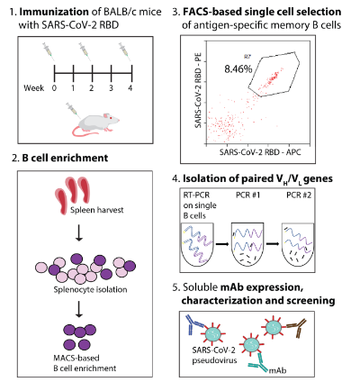
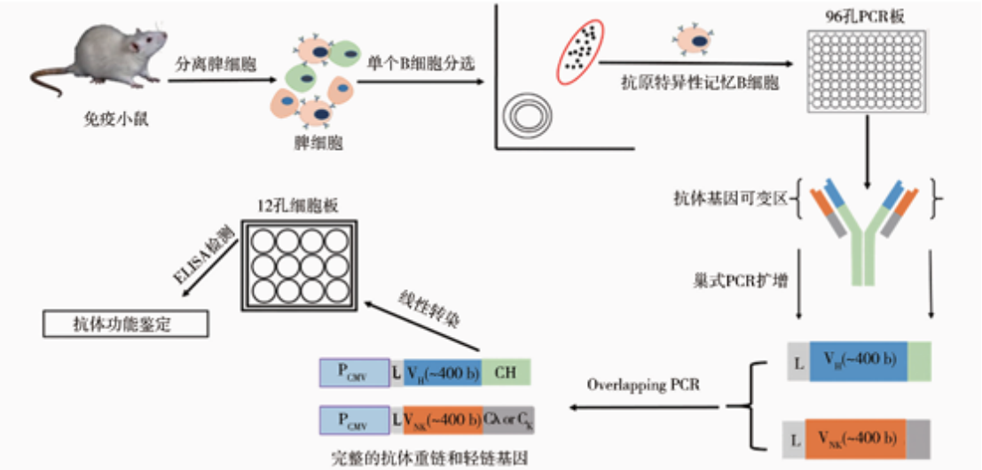
ELISA, Western blotting or immunoprecipitation are used to screen high-valence serum samples. For situations where monoclonal antibodies need to be generated, B cells can be isolated from immunized alpacas, single-cell screening can be performed, and then specific antibody genes can be cloned using molecular biological methods (such as PCR, plasmid construction, etc.). If needed, the affinity of the resulting antibody can be enhanced by in vitro affinity maturation techniques (e.g., phage-based or yeast-based display techniques).

Production and validation of high-affinity antibodies

Use of suitable expression systems (e.g. mammalian cells, E. coli, etc.) to produce recombinant antibodies. Antibodies are purified and thoroughly analyzed for biological function and binding properties to ensure that they have the expected high affinity and specificity.


References

[1] Cortez-Retamozo V .Efficient Cancer Therapy with a Nanobody-Based Conjugate[J].Cancer Research, 2004, 64(8):2853-2857.DOI:10.1158/0008-5472.CAN-03-3935.

[2] Meyer T D , Muyldermans S , Depicker A .Nanobody-based products as research and diagnostic tools[J].Trends in Biotechnology, 2014, 32(5).DOI:10.1016/j.tibtech.2014.03.001.

[3] Steyaert J , Kobilka B K .Nanobody stabilization of G protein-coupled receptor conformational states.[J].Current Opinion in Structural Biology, 2011, 21(4):567-572.DOI:10.1016/j.sbi.2011.06.011.

176 月/24
VHH

Production of Nanobody by Immunizing Alpaca

Background Introduction

Antibodies are biological protein molecules produced by immune B cells stimulated by antigens that can bind specifically to antigens. Due to its ability to bind antigens with high specificity and affinity, antibodies are widely used in academic research, disease diagnosis, and various aspects of medical drugs[1].

A conventional antibody molecule (IgG) is a rather conserved protein molecule consisting of two identical heavy chains and two identical light chains. The light chain of an antibody contains one VL region and one CL region, while the heavy chain has one VH region and three CH regions (CH1, CH2, and CH3).

VH region and VL region together constitute the smallest unit of antigen recognition by traditional antibodies, and the sequence difference of antibody variable region determines that antibodies can specifically recognize different antigens[2]. On the other hand, the CL region and CH region are relatively conserved and known as the constant region of antibodies, in which the CH2 and CH3 regions of the CH region play an important role in recruiting immune cells for ADCC and CDC functions.

Heavy Chain antibodies are special antibodies naturally occurring in camels and cartilaginous fish that are composed of only two heavy chain antibodies in addition to traditional antibodies and contain only one Variable Domain of Heavy Chain Antibody (VHH) and two conventional CH2 and CH3 regions, with the absence of CH1 region. Heavy chain antibodies bind to antigens via a variable region (VHH) on the heavy chain that can be stably present alone in vitro and are called camel single domain nanobody (SdAb) or nanobody. The nanobody crystal is 2.5nm wide and 4nm long, and the molecular weight is only 1/10 (about 15kD) of the traditional complete antibody, but it still has complete antigen recognition ability, and the VHH sequence is generally obtained by phage screening.

Thanks to the tiny structure, complete antigen recognition ability, and phage screening technology, the complete VHH sequence can be obtained, and the nanobody can be mass-produced by recombinant expression in vitro, effectively avoiding the problem of batch-to-batch difference of traditional antibodies.

Compared with traditional antibodies, nanobodies have small molecular weight and simple structure. Due to the advantages of small molecular weight, nanobodies have many features, which makes nanobody show great potential in new drug discovery[3] : They have stronger specificity in binding to targets, and can bind to sites that traditional antibodies cannot bind to; Higher tissue penetration; Higher stability such as high temperature resistance; Suitable for industrial large-scale production; Easier to transform and optimize; Easier to humanize.

Because of these characteristics of nanobody, more and more research institutions and drug manufacturers pay attention to and try to use nanobody in different scenarios. The development of nanobody is different from the traditional monoclonal antibody production method by hybridoma, which generally selects candidate nanobody by immunizing alpaca, constructing phage library and displaying phage, and then carries out the verification experiment of binding to antigen after expression and purification of nanobody.

VHH

Figure 1:The difference between conventional antibody and nanobody

Introduction to Immunogen Types

At present, the acquisition of single-domain antibodies is generally achieved by immunizing alpaca and maturing the antibodies of the immune system itself in alpaca. B lymphocytes are separated, RNA is extracted, cDNA is obtained by reverse transcription, cDNA is used as the substrate for PCR amplification to obtain diversified nanoparticle antibody gene fragments, and then the diversified nanoparticle antibody gene fragments are connected to phage granules to construct phage library. Then, the most suitable antibody was selected from the alpaca antibody library by phage display screening technology and the nanobody was verified. The whole process mainly includes alpaca immunization, phage library construction, antibody screening, expression purification and verification.

Antibody Discovery-KMDBioscience

Figure 2:The difference between conventional antibody and nanobody

https://www.kmdbioscience.com/article/Generation-Process-of-Nanobody.html

The immunogens used to immunize alpacas include:

Protein immunogen

Protein immunogen is generally composed of whole proteins or recombinant protein fragments, which can well simulate the epitope of the antigen under natural conditions, stimulate a strong immune response, and thus induce the production of nano antibodies with high affinity and specificity.

Small molecule immunogen

Small molecule usually refers to the molecular weight of less than 1 kDa compound. Due to its low molecular weight, it is not sufficient on its own to trigger a response from the immune system, so it needs to be coupled with a larger carrier protein to enhance its immunogenicity.

Polypeptide immunogens

Polypeptide immunogens are composed of short chain amino acids, can be linear or conformationally qualified, and are usually used to simulate local epitopes of protein antigens, which need to be properly designed and modified to improve their stability and immunogenicity.

Virus immunogen

Viral immunogens are specific proteins or virus particles extracted from viruses that induce an immune response from the host. These immunogens are usually inactivated, unable to cause disease, but sufficient for the host’s immune system to produce a defense against the virus.

DNA immunogen

This type of immunogen is usually prepared by synthesizing the coding DNA sequence of the target protein. The advantage of DNA immunogen is that it can precisely control the coding sequence of proteins, enabling the production of specific antibodies. In addition, DNA immunogen preparation is relatively simple and cost-effective.

RNA immunogen

RNA immunogen is very useful for studying specific RNA subtypes and post-transcriptional modifications. However, it is difficult to maintain its stability and complex to operate.

Cell line type Immunogen

This is a method that uses the cell line as the immunogen, usually through the expression of the target protein by the cell line to produce antibodies.

Immunization Technique

The selection of alpaca and the immune antigen are the key to the success of immunization. Choose healthy and strong, good mental state, moderate size of the alpaca is blank alpaca. The purity of the immune antigen and its correct conformation are crucial to the screening of suitable antibodies after immunizing alpacas for subsequent use, and the purity of the protein antigen is generally not less than 90%.

lymphocyte separation: Timely cell separation can effectively prevent hemolysis after blood collection to achieve the best separation effect.

The choice of immune cycle can affect the immune effect, according to experience, 1-2 weeks of immunization interval can make alpacas have a good immune response to most antigens.

References

[1] Cortez-Retamozo V .Efficient Cancer Therapy with a Nanobody-Based Conjugate[J].Cancer Research, 2004, 64(8):2853-2857.DOI:10.1158/0008-5472.CAN-03-3935.

[2] Meyer T D , Muyldermans S , Depicker A .Nanobody-based products as research and diagnostic tools[J].Trends in Biotechnology, 2014, 32(5).DOI:10.1016/j.tibtech.2014.03.001.

[3] Steyaert J , Kobilka B K .Nanobody stabilization of G protein-coupled receptor conformational states.[J].Current Opinion in Structural Biology, 2011, 21(4):567-572.DOI:10.1016/j.sbi.2011.06.011.

146 月/24

Stable cell line construction technique common problems and solutions-KMD Bioscience

What is the difference between transient and stable cell lines?

Transient cell lines are those in which the target gene is not permanently integrated into the host cell genome, resulting in gene expression occurring only for a limited period. Stable cell lines contain target genes that are integrated into the host genome to support the long-term expression of target genes.

Is the time and expense necessary to build a stable cell line?

If trial and error is required on cells that overexpress or reduce the expression of the desired gene, then the construction of stable cell lines is necessary. First, it can reduce the difference in experimental results caused by the difference in transfection efficiency in different batches of experiments, and improve the repeatability of the experiment. Second, after the construction of the cell line, there is no need to perform instantaneous transfection experiments and repeated preparation of plasmids, reducing the workload. Thirdly, expensive transfection reagents are no longer used to save experimental costs.

What are the advantages of stable cell lines?

Key advantages of stable cell lines include significantly reduced variability between production batches, applicability to a wide range of research and production applications, and robust growth performance.

How many generations can the target gene be stably expressed?

The characteristics of the lentiviral system determine that the target genes are always expressed like the cell’s own genes, and are not lost in the process of passage.

What is the difference between monoclonal and polyclonal cell lines?

Monoclonal cells are cloned and screened after transfection of the target gene. The cell line formed by the expansion of one cell has the same integrated location and copy number of the target gene, and the expression level can be maintained for a long time. Polyclonal cells were directly screened by a drug resistance gene after transfection of target gene. It’s a mixture of multiple positive cell clones. For information on monoclonal and polyclonal cell lines, see our monoclonal cell Lines Introduction.

How long does it take to produce a stable cell line?

Developing a stable cell line typically takes 6 to 12 months, and this time frame varies depending on the complexity of the specific target protein or biologics being pursued.

Can stable cell lines be constructed from all cell lines?

Not all cell lines can be constructed into stable cell lines. Two conditions need to be met to construct stable cell lines: 1. Stable cell passage; 2. The transfection efficiency of cells met the requirements. We offer a free pre-lab service to test whether your cell line can be used for stable cell line construction.

How to choose the right lentiviral vector?

The selection of lentiviral vectors mainly requires consideration of promoter selection, whether fluorescent protein tags are required, and whether fluorescent protein tags are required for fusion expression. Commonly used lentiviruses are pLV-puro, pLV-EGFP-C, pLV-sfGFP (2A) puro.

What is the method for constructing stable cell lines?

Stable cell lines were constructed by lentivirus transfection. The characteristic of this system is that the target gene is first integrated into the genome and then expressed, without instantaneous expression process. Therefore, the screened cells are stable and integrated.

References

[1] Tihanyi B, Nyitray L. Recent advances in CHO cell line development for recombinant protein production. Drug Discov Today Technol. 2020 Dec;38:25-34.

[2] Chen LQ, Wang YN, Li DY, Xu LY, Li LJ, Li ZH, Liu H, Liu Q. Construction of a Stable Expression Cell Line of Human Phospholamban. Fa Yi Xue Za Zhi. 2021 Oct 25;37(5):615-620.

[3] Lü X, Zhou Z, Zhu L, Zhou J, Huang H, Zhang C, Liu X. [Construction and identification of a HEK293 cell line with stable TrxR1 overexpression]. Nan Fang Yi Ke Da Xue Xue Bao. 2022 Apr 20;42(4):554-560.

136 月/24

The Role of Single B Cell Screening in Vaccine Development-KMD Bioscience

Monoclonal antibodies play a key role in the fight against the novel coronavirus and effectively reduce the progression of mild to severe symptoms in patients. However, future progress in this field depends on the timing and technology of antibody development. A recent article published in the journal Nature-Scientific Report demonstrates the use of single B cell clonal screening for rapid and reliable identification of high-affinity, potent neutralizing antibodies to the novel coronavirus (SARS-CoV-2) and explains how to enhance neutralizing activity.

After vaccination, different types of antibodies are generally produced, taking neutralizing antibodies as an example: neutralizing antibodies are antibodies produced by the organism stimulated by the outermost envelope or capsid antigen of the virus that can bind to the virus and make it lose its infectiousness. In the current research on COVID-19 vaccine, neutralizing antibodies belong to the focus of research, such as the receptor-binding domain (RBD) for S1 protein and the N-terminal domain (NTD) for S1 protein[1].

The neutralizing antibody studied in this study belongs to the former. For the S1 protein receptor binding domain (RBD), it can bind to the RBD region of SARS-CoV-2 and block the binding of this region to the ACE2 receptor, so as to prevent the virus from invading cells. This type of antibody is the most produced by the human body and the most important type of antibody that has a good effect on blocking the virus.

Antibodies can be found faster through single B cell screening. The antibody discovery process can be shortened to a few weeks through single B cell sorting. This pioneering approach involves isolating live cells from immunized animals or rehabilitated patients, classifying antigen-specific cell subpopulations, and recovering paired VH/VL antibody genes by RT-PCR and PCR; The encoded antibodies can be expressed and characterized to identify good performing clones, as shown in Figure 1.

Figure 1: A review of methods for isolating and identifying effective SARS-CoV-2 neutralizing antibodies from single B cells

The advantage of this method is that it uses the native antibody sequence with high somatic mutation and preserves the native VH/VL pairing. In addition, the discovery process is high-throughput, allowing further evaluation of the highly immune antibody library.

To evaluate the effectiveness of single B cell screening in isolating neutralizing antibodies against SARS-CoV-2 virus, we prepared a recombinant form of SARS-CoV-2 receptor-binding domain (RBD) in HEK293T cells and purified its protein[2].

After immunizing BALB/c mice, anti RBD IgG1+B cells were isolated from spleen material by single cell sorting, and the VH /VL gene was treated. The resulting VH/VL gene pairs were cloned into mammalian expression plasmids (including the constant region of human IgG1 and the kappa light chain region for chimeric antibody expression) and transfected into HEK 293-6E cells instantaneously. Antibody identification includes the evaluation of Protein A magnetic beads’ purification, antigen-binding ability, neutralizing activity and other properties.

After the recombinant antibody is produced, its affinity must be analyzed for downstream applications. Antibody affinity detection methods include biofilm interferometry (BLI), solid phase radioimmunoassay (SP-RIA), equilibrium dialysis, binding antigen precipitation, radioimmunoassay (RIA), ELISA, and surface plasmon resonance (SPR).

RBD regions of SARS-CoV or SARS-CoV-2 with His labels were incubated separately with Dynabeads™ magnetic beads to detect the specificity of neutralizing antibodies. The two magnetic beads were incubated separately with different concentrations of antibodies, and the unbound magnetic beads were eluted. It was then incubated with Alexa Fluor®488 fluorescent secondary antibody (goat-derived, anti-human IgG, Fc fragment specific affinity purified secondary antibody, Jackson ImmunoResearch) and analyzed by flow cytometry.

B-cell screening has been shown to rapidly identify high-affinity, potent SARS-CoV-2 neutralizing antibodies. In addition, its use in combination with antibody modification is expected to be an effective strategy to prevent COVID-19.


References

[1] Davis-Marcisak E F , Deshpande A , Stein-O’Brien G L ,et al.From bench to bedside: single-cell analysis for cancer immunotherapy[J].Cancer Cell, 2021, 39(8).DOI:10.1016/j.ccell.2021.07.004.

[2] Zhang Z, Xu Q, Huang L. B cell depletion therapies in autoimmune diseases: Monoclonal antibodies or chimeric antigen receptor-based therapy? Front Immunol. 2023 Feb 10;14:1126421. doi: 10.3389/fimmu.2023.1126421. PMID: 36855629; PMCID: PMC9968396.

126 月/24

Single B Cell Sequencing and Antibody Genomics-KMD Bioscience

Antibody is an important component in biological drugs and has many application prospects. However, traditional antibody research methods often only provide information at the global level and cannot reveal the heterogeneity between different cells. With the development of single-cell sequencing technology, researchers can gain a deeper understanding of the expression and function of antibodies at the level of individual cells, thus providing more comprehensive information for antibody research and development. This paper will explore the application of single-cell sequencing technology in antibody research and analyze its potential in antibody discovery, optimization, and therapeutic monitoring.

Introduction to Single B Cell Sequencing

Single-cell sequencing is a technique that enables high-throughput genome, transcriptome, or proteome sequencing of a single cell. It enables efficient sequencing of intracellular molecules by isolating, trapping, and placing individual cells into micro-reactors. Currently, common single-cell sequencing technologies include single-cell RNA sequencing (scRNA-seq), single-cell DNA sequencing (scDNA-seq), and single-cell proteomics sequencing.

Figure 1: Flow chart of single-cell sequencing

Significance and application of single cell antibody sequencing

Analysis of immune diversity: Single cell sequencing technology can help researchers conduct a comprehensive analysis of the diversity of antibodies. By performing RNA-SEQ on a single B cell, it is possible to obtain transcript information of its antibody genes, so as to understand the diversity and variation of antibodies in different cells. This helps researchers better understand the structure and function of antibodies, providing guidance for antibody design and optimization.Single cell antibody sequencing technology reveals the diversity and variation of antibodies in the immune system, which can help scientists understand the specificity and diversity of different cells in the immune response, and thereby reveal the working principle of the immune system and the mechanism of disease development.

Disease diagnosis and treatment strategies: Single-cell antibody sequencing technology can help doctors diagnose and treat diseases more accurately. By analyzing the antibody sequences in individual cells of patients, we can understand the characteristics of disease development and the variation of antibodies, so as to provide a basis for the development of personalized treatment strategies.

Biologic drug development and optimization: In the field of biologic drugs, single-cell antibody sequencing technology can help scientists better understand the differences in drug mechanisms of action and efficacy. By analyzing antibody sequences in a single cell, drug candidates with high affinity and specificity can be screened and their performance and effects can be further optimized.

The development of single-cell antibody sequencing technology has revealed the diversity and complexity of the immune system, bringing revolutionary progress to immunology and biologic drug research. However, the technology still faces some challenges, such as sequencing errors and the complexity of data analysis. Future research should aim to improve the accuracy and efficiency of the technique and further explore the potential of single-cell antibody sequencing.

The emergence of single-cell antibody sequencing technology has revealed the mystery of immune diversity for us, breaking through the limitations of traditional antibody sequencing. Through this technology, we can deeply understand the immune characteristics of each individual cell, providing a more accurate method for the diagnosis and treatment of diseases, while providing new tools and ideas for the development and optimization of biological drugs. As technology continues to evolve and innovate, it is reasonable to believe that single-cell antibody sequencing technology will play an increasingly important role in future research.

References

[1] Yang XW, Sun K. Research progress of single cell sequencing in the diagnosis and treatment of hematological diseases. Zhonghua Xue Ye Xue Za Zhi. 2019 May 14;40(5):443-446. Chinese. doi: 10.3760/cma.j.issn.0253-2727.2019.05.020. PMID: 31207715; PMCID: PMC7342236.

[2] Busse, C.E., Czogiel, I., Braun, P., Arndt, P.F. & Wardemann, H. Single-cell based high-throughput sequencing of full-length immunoglobulin heavy and light chain genes. Eur J Immunol 44, 597-603 (2014).

[3] Setliff I, Shiakolas AR, Pilewski KA et al. High-ThroughputMapping of B Cell Receptor Sequences to Antigen Specificity Cell. 2019 Dec12;179(7):1636-1646.e15.

116 月/24

Single B Cell Screening in Autoimmune Disease Research-KMD Bioscience

Autoimmune disease is a disease caused by the body’s immune system being mistakenly directed to attack the host itself, and more and more patients with autoimmune diseases need treatment. Researchers have now described more than 80 autoimmune diseases, which can be systemic, such as systemic lupus erythematosus(SLE), which affects the skin, joints, kidneys and central nervous system, or organical, such as type 1 diabetes, which primarily affects the body’s pancreas. Loss of B cell or T cell tolerance is often associated with autoimmunity. In recent decades, the treatment of autoimmune diseases has shifted from the use of hormones and conventional immunosuppressive drugs to the use of biologics. The proliferation and maturation of B lymphocytes play an important role in the pathogenesis of autoimmune diseases. Single B cell screening is becoming increasingly important in the treatment of autoimmune diseases. Monoclonal antibodies that target B cells and plasma cells can effectively treat a wide range of autoimmune diseases, underscoring the importance of B cells in the pathogenesis of such diseases.

In autoimmune diseases, B-cell-associated monoclonal antibodies mainly target CD20, CD19, CD22, CD38 and B-cell activating factor (BAFF). Some drugs have been approved by the US Food and Drug Administration, such as rituximab, Beliuzumab, orfathomumab, etc.

Antibodies targeting CD20 are currently the most widely used monoclonal antibodies, and although these antibodies have the same target, their structures and indications are quite different. The first generation of anti-CD20 drugs, such as rituximab, has good efficacy in pemphigus, rheumatoid arthritis, granulomatous polyvasculitis and microscopic polyvasculitis. Second-generation anti-CD20 drugs, including ocrelizumab, Oftolumab, and veltuzumab (not approved), contain both humanized and fully human monoclonal antibodies to reduce immunogenicity and extend the half-life of the drug. These drugs can bind Fc receptors on B cells more closely. The approved ocrelizumab and oftulizumab are mainly used in the treatment of multiple sclerosis. The third generation of anti-CD20 monoclonal antibodies, represented by Ortuzumab, has a different binding epitope from rituximab and does not induce CD20 aggregation or antibody internalization, so it is more effective and less resistant.

Inellizumab is a humanized anti-CD19 monoclonal antibody that has been approved by the FDA for the treatment of neuromyelitis spectrum disorder (NMOSD). NMOSD is a rare recurrent autoimmune disease of the central nervous system that can lead to paralysis and blindness. Beliuzumab is a fully human anti-BAFF monoclonal antibody that rapidly reduces naive B cells and B cells in early developmental stages, and currently treats SLE and lupus nephritis. In addition, there are several drugs that target other B cell surface antigens, such as epratuzumab, which targets CD22, and daratumumab, which targets CD38, that are being tried for the treatment of SLE. SLE is characterized by the reaction of B cells with autoantigens to produce autoantibodies. These autoantibodies may appear years before the clinical onset of SLE. Overproduction of autoantibodies leads to an inflammatory cascade and a massive immune response that ultimately leads to organ damage in the patient. In addition to secreting autoantibodies, B cells also secrete cytokines that promote inflammation.B cells and plasma cells (effector B cells) play an important role in the pathogenesis of SLE, and their associated antigens are the main therapeutic targets, such as CD19, CD20, CD40, etc. (Figure 1).

Figure 1: The maturation process of B cells


References

[1] Davis-Marcisak E F , Deshpande A , Stein-O’Brien G L ,et al.From bench to bedside: single-cell analysis for cancer immunotherapy[J].Cancer Cell, 2021, 39(8).DOI:10.1016/j.ccell.2021.07.004.

[2] Zhang Z, Xu Q, Huang L. B cell depletion therapies in autoimmune diseases: Monoclonal antibodies or chimeric antigen receptor-based therapy? Front Immunol. 2023 Feb 10;14:1126421. doi: 10.3389/fimmu.2023.1126421. PMID: 36855629; PMCID: PMC9968396.

[3] Arbitman L, Furie R, Vashistha H. B cell-targeted therapies in systemic lupus erythematosus[J]. J Autoimmun. 2022 Aug 10:102873. doi: 10.1016/j.jaut.2022.102873. Epub ahead of print. PMID: 35963808.

076 月/24

Overview of Single B Cell Screening Technology-KMD Bioscience

Development of Single B Cell Screening Technology

In 1975 Milstein[1] first described a method to produce monoclonal antibody by hybridoma technology, which greatly promoted the development of immunology. Monoclonal antibody has become an important scientific research tool and therapeutic molecule, and this technology is still the main technology for the preparation of monoclonal antibody. It plays an important role in cell biology technology. Monoclonal antibody is an important tool in the field of medicine and immunology, and plays a great role in the pathogenesis, diagnosis and treatment of pathogens. At present, the most commonly used methods of monoclonal antibody preparation are hybridoma technology and phage display technology. Hybridoma technology is the fusion of spleen cells and bone marrow cells of immune mice to form hybridoma cells that survive in vitro for a long time and secrete immune proteins, and then produce monoclonal antibodies targeting the same antigen binding site, which is the most classic monoclonal antibody preparation technology. This method is mature and transparent, and can produce strong specific monoclonal antibodies, but the preparation time is long, the repetitive production performance is poor, the difference between antibody batches is large, and hybridoma cells are very easy to appear in the culture process chromosome loss, clonal competition, hybridoma decreased ability to secrete antibodies, etc., increase the risk of antibody development. Phage display technology is to insert variable region genes of antibodies into phage genes, display the expressed antibodies to the surface of phage, construct phage display antibody library, simulate the production of antibodies in vitro, and screen out antibodies against different antigens. The phage display method does not need to immunize animals, shortens the antibody preparation cycle, and has a large screening capacity, which can screen hundreds of millions of clones in a short time. However, the random combination of antibody variable region genes will lead to the loss of natural pairing of antibody heavy chain and light chain, and the modification of antibody is different from that of mammals, so the application of this method in production is limited.

In recent years, the single B cell antibody screening technology has been gradually developed, and then it has been applied to the preparation of monoclonal antibodies. This technology uses flow cell sorting to fluorescentially label specific antigens, and uses the principle that antigens can specifically bind to the BCR on the surface of B cells to screen out single target B cells. Then, the heavy chain and light chain genes of antibodies are obtained by PCR amplification, and the antibody genes are constructed on the expression vector. Transfected into eukaryotic cells to express monoclonal antibodies(Figure 1). Different from traditional hybridoma cell sorting technology, single cell screening technology can directly isolate, culture, analyze and screen single B cells, so as to accurately and efficiently screen out B cells secreting target antibody molecules, and then obtain the target antibody sequence by combining single cell sequencing technology. This technique can quickly produce monoclonal antibodies against humans, rats, rabbits, alpacas, pigs, chickens, dogs and other animals. The method is time-saving and efficient, and retains the natural pairing of antibodies, and has been widely used in the research of infectious diseases such as HIV, COVID-19 , and influenza.

Figure 1: Schematic diagram of preparation of McAbs based on single B cell antibody technology[2]

Significance of Single B Cell Screening Technology inbiotechnology research

Monoclonal antibody plays an important role in the prevention, diagnosis and treatment of diseases, especially in the study of immune mechanism. With the development of monoclonal antibody preparation technology, single B cell antibody preparation technology has become a new generation of rapid monoclonal antibody preparation method. Compared with traditional antibody preparation technology, this technology has the advantages of fast, efficient and high yield, and the antibody expressed has a natural conformation, which can not only be used for the development of antibodies related to pathogenic microorganisms and the study of the mechanism of virus cross-species transmission, but also play an important role in anti-tumor therapy and anti-autoimmune diseases.

Categories Single B Cell Technique Hybridoma Technology Phage Display Technology
Antibody Natural Natural Unnatural
Antibody Affinity High High Medium
Druggability High High Low
Time 16 to 20 Weeks, Optional Rapid Immunization 20 to 26 Weeks, Optional Rapid Immunization 16 to 20 Weeks, Optional Rapid Immunization
Antibody Gene Sequence Direct Acquisition Further Sequencing Required Direct Acquisition
Antibody Diversity High Medium Medium
The species that can be screened Unrestricted Species Mouse and Rabbit Unrestricted Species

References

[1] Köhler, Milstein C .Pillars Article: Continuous cultures of fused cells secreting antibody of predefined specificity. Nature, 1975, 256 (5517): 495–497.[J].Journal of Immunology Official Journal of the American Association of Immunologists, 2005, 174(5):2453-2455.

[2] CHEN Yang, LIU Tong, ZHANG Jia-qi, LIAO Hua-xin, LIN Yue-zhi, WANG Xiao-jun, WANG Ya-yu. Screening of Monoclonal Antibodies Targeting the Equine IgG1 Based on Single B Cell Antibodies Gene Amplification Technology. China Biotechnology, 2022, 42(4): 17-23.

066 月/24

Monoclonal vs. Polyclonal Antibodies: The Impact of B Cell Screening-KMD Bioscience

When pathogens (such as bacteria, viruses, etc.) invade the body, B cells secrete antibodies (also known as immunoglobular proteins) to neutralize the antigen. An antibody protein is a Y-shaped molecule consisting of four polypeptide chains (two heavy chains and two light chains). The Y-shaped structure contains an antibody binding site (paratope, which can be interpreted as a “lock”) at each of the two tips. Each antibody binding site can specifically bind an antigen-epitope (which can be understood as a “key” to a “lock”)[1]. The unique and specific correspondence between antibody-binding epitope and antigen-epitope is the basis of the high specificity of antibody-antigen binding. Antibodies are widely used in pharmaceutical research and development. There are two main classes of antibodies: monoclonal antibody (Mabs) and polyclonal antibody (Pabs)[2].


Figure 1: Antibody structure


Polyclonal antibodies are mixtures of antibodies produced by different B cells in the body against the same antigen. They can recognize and bind different epitopes on that single antigen. Polyclonal antibodies are produced by injecting certain immunogens into animals. After animal immunization, polyclonal antibodies can be obtained directly from serum (blood after removal of coagulation proteins and red blood cells) or further purified to remove other serum proteins. The specific process includes preparing antigens, immunizing animals, collecting serum after qualified titer determination, purifying and identifying antibodies. The process is simple, low cost and fast[3].

Figure 2: Method of production of polyclonal antibodies

Monoclonal antibodies are produced by identical B cells (clones of the same mother cell). This means that monoclonal antibodies can only recognize a specific single epitope. Unlike polyclonal antibodies, which are produced directly by animals, monoclonal antibodies are produced using tissue culture techniques in vitro. The process is as follows: ① Animal immunity and PBMC isolation. Cows are selected as experimental animals and are immunized 4-5 times by injecting a target antigen (usually a target protein or polypeptide) to stimulate an immune response, thereby activating the cow’s immune system and causing B cells to produce antibodies. Then ELISA assay, peripheral blood collection and PBMC isolation were performed. ② Flow sorting. Antigen specific single B cells were sorted by PBMC flow sorting method. ③ Single B cell culture screening. The single B cells were first cultured in 5-20 plates with 96-well plates in vitro, and then the supernatant of the cell culture was screened by ELISA or pre-interventional detection such as FACS. ④ Sequencing of positive clones. At least 20 positive cell clones were selected for RT-PCR to amplify their antibody variable region genes and then sequenced. ⑤ Vector construction, antibody expression and purification. Finally, the mammalian expression vector, cell transfection and ELISA were verification.

Figure 3: Method of production of monoclonal antibodies

Figure 4: The mode of binding of (a) polyclonal antibody to (b) monoclonal antibody to antigen

Monoclonal antibody and polyclonal antibody have their own distinct characteristics and advantages. The specificity of monoclonal antibody is high, and once successfully prepared, both permanent monoclonal antibody and multi-clonal antibody have their own distinct characteristics and advantages. Monoclonal antibody has a high specificity, and once it is successfully prepared, it can sustainably produce completely consistent monoclonal antibody, so its specificity can be comprehensively and systematically verified. However, if the identified epitope is destroyed, the results of the experiment will be greatly affected, which is also one of the disadvantages of monoclonal antibodies. However, the specificity of polyclonal antibodies is poor, even if the same antigen is used to prepare polyclonal antibodies, there will be differences between different batches, so there are great limitations in specificity and consistency. Therefore, when using multiple antibodies for immune detection, it is easier to create a background, such as a mixed band in WB, a darker background in IHC, and so on. Although there is still a crossover. However, because multiple antibodies recognize multiple epitopes, even if a few epitopes are destroyed or the antigen conformation is changed, the results of the experiment will not be affected. Under the same conditions, the use of multiple antibodies can improve the sensitivity of the detection, and it is easier to detect proteins with low abundance.

References

[1] Lipman NS, Jackson LR, Trudel LJ, Weis-Garcia F. Monoclonal versus polyclonal antibodies: distinguishing characteristics, applications, and information resources. ILAR Journal. 2005;46(3):258-268. doi:10.1093/ilar.46.3.258

[2] Bregenholt S, Jensen A, Lantto L, Hyldig S, Haurum JS. Recombinant human polyclonal antibodies: a new class of therapeutic antibodies against viral infections. Curr. Pharm. Des. 12(16), 2007–2015 (2006). doi: 10.2174/138161206777442173

[3] Grodzki AC, Berenstein E. Antibody purification: ammonium sulfate fractionation or gel filtration. Methods MolBiol. 2010;588:15-26. doi:10.1007/978-1-59745-324-0_3

056 月/24
crisper

Integrating Single B Cell Screening with CRISPR Technology-KMD Bioscience

Single-cell CRISPR Screening

CRISPR screening (CRISPR-Screen) technology quantifies the changes of sgRNA targeting target genes before and after screening based on sequencing means and is currently widely used to screen candidate genes associated with specific functional phenotypes such as cell growth, differentiation, immune tolerance, or drug resistance. Although CRISPR screening can study gene function to a certain extent, this technology still has great limitations in studying the regulatory mechanisms downstream of the target gene.

Single-cell CRISPR screening is a powerful way to study underlying developmental, disease, and therapeutic response mechanisms. By directly correlating CRISPR perturbations and single-cell gene expression data cell by cell, it is possible to analyze hundreds of different CRISPR perturbations and detect single guide Rnas (Sgrnas) with direct gene expression phenotypes in hundreds to tens of thousands of cells without knowing the cell type or markers.

10x single-cell sequencing combined with Feature Barcode technology enables simultaneous detection of CRISPR perturbations and the resulting multiple transcriptional signatures, greatly expanding the operability, scalability, and resolution of high-throughput functional screening, allowing us to further explore the mysteries of biology.

Traditional CRISPR hybrid screening can provide limited information about how a gene affects other genetic pathways, and also requires trade-offs between depth and scale of characterization. For example, when we have multiple genes of interest and want to study all the targets at once, if we use traditional CRISPR hybrid screening, we can directly mix together Sgrnas designed for these target genes and transfect them into cells, and we will end up with a group of cells that we do not know what has been transfected. The single-cell CRISPR screen is a very effective alternative to this problem, not only screening thousands of GRnas in a single experiment, but also obtaining perturbed full transcriptome data at the same time to provide the clearest understanding of cell type-specific gene function and pathway analysis.

Experimental procedures and principles of 10x single-cell CRISPR screening

Single-cell CRISPR screening requires first designing gRNA according to target gene, then assembling the designed gRNA sequence into lentiviral vector for subsequent cell transformation, then transfecting the cells with the assembled lentivirus, and screening the transfected cells by flow or antibiotic method.

Finally, the screened cells were captured through the 10x platform: In the oil-in-water reaction system, the oligo sequence in Gel Beads will capture the gRNA sequence in the cells, and the protospacer sequence on the gRNA will know which gRNA is transferred into the cells. In this way, the cell expression profile data can be examined at the same time to detect which gRNA is edited by the cell and the gene expression after editing.

crisper

Figure 1: Experimental procedures of single-cell CRISPR screening

Application of 10x single-cell CRISPR screening

Single-cell CRISPR screening directly correlates gRNA within the same cell with full transcriptome gene expression through sequencing, helping us to fully understand cell type-specific gene function and opening up a new era of biological exploration. As a cutting-edge technology, single-cell CRISPR screening will gradually be widely used in many important research fields such as immunology, neuroscience, and cell development.

With the depth of information brought about by single-cell CRISPR screening methods, researchers are able to gain insight into disease mechanisms and new pathways for therapy development, not only for neurodegenerative diseases, but for all major types of disease. Gene function itself is no longer a simple reading of cell death or survival. Instead, technological innovations in CRISPR screening allow our experiments to match the true complexity of biology, resolving the perturbed transcriptomic effects at single-cell resolution, resulting in a rich picture of gene function in specific cell populations.

References

[1] Wu C A M , Roth T L , Baglaenko Y ,et al.Genetic engineering in primary human B cells with CRISPR-Cas9 ribonucleoproteins[J].Journal of Immunological Methods, 2018:S0022175918300073.DOI:10.1016/j.jim.2018.03.009.

[2] Rodríguez-Pinto, D. B cells as antigen presenting cells. Cell. Immunol. 238, 67–75 (2005).

[3] Amanna, I. J. & Slifka, M. K. Mechanisms that determine plasma cell lifespan and the duration of humoral immunity. Immunol. Rev. 236, 125–138 (2010).

046 月/24

Ethical Considerations in Single B Cell Research-KMD Bioscience

As a new generation of antibody development technology, single B cell antibody technology can efficiently and rapidly isolate antibodies from single B cells, with outstanding advantages such as high specificity, high activity, and high affinity. However, when researching single B cells, we must face a range of ethical issues and ethical guidelines. Ethical considerations are essential in biological research to ensure responsible and humane treatment of living organisms, respect for the rights of participants, and the integrity of scientific inquiry.

Ethical issues of using live animals as research subjects

The use of live animals in research is subject to ethical principles and government regulations to ensure humane treatment, welfare and ethical considerations regarding their use. Here are some of the key principles and regulations that guide the use of live animals as research subjects:

Three R

The Three R’s are the basic ethical framework for animal research, emphasizing the principles of replacement, reduction, and refinement. It encourages researchers to seek alternatives to animal testing where possible (alternatives), minimize the number of animals used (reduction), and improve experimental procedures to minimize the pain, suffering, and suffering experienced by animals (improvement).

Institutional Animal Care and Use Committee (IACUC)

The IACUC is established in research institutions to review and oversee all research protocols involving animals. These committees ensure that research projects comply with ethical guidelines and regulations, and assess the scientific and ethical justifications for animal use. IACUC reviews and approves research protocols, monitors animal welfare and enforces compliance with relevant regulations.

Animal welfare regulations

Most countries have specific regulations and guidelines regarding the use of animals in research. These regulations typically outline standards for animal care, housing, nutrition, veterinary care, and handling procedures. They also include provisions for pain management, euthanasia, and humane endpoints for animal testing. Researchers must comply with these regulations to ensure the welfare and well-being of the animals used in research.

Species-specific guidelines

Animals of different species may require special consideration in research. Researchers must understand the specific biological and behavioral needs of the animals they study. Species-specific guidelines provide additional advice on how animals are kept, enriched, handled and cared for to ensure their health and minimize stress.

Anesthesia, analgesia, and euthanasia

When animals undergo surgery that may cause pain or suffering, researchers need to provide appropriate anesthesia and analgesia to minimize suffering. Euthanasia methods should be humane, ensuring a quick and painless death when necessary.

Record keeping and reporting

Researchers are required to maintain detailed records of the animals used in the study, including their origin, feeding conditions, health status, procedures performed, and any adverse events. This information is essential to ensure transparency, track animal welfare and provide accountability.

Ethical review and approval

Researchers are usually required to submit research protocols involving animals to IACUC or an equivalent ethics committee for review and approval. The Committee assesses the scientific and ethical justifications for the use of animals and assesses proposed procedures to ensure that they meet the necessary standards for animal welfare and ethical considerations.

Ethical issues of data processing and release

Data processing and publication is an important part of cell biology research. When processing data, researchers need to adhere to the principles of scientific integrity and data integrity to ensure the authenticity and reliability of the data. Researchers should disclose any potential conflicts of interest that could bias their research or affect its objectivity. This includes financial interests, professional relationships or personal relationships that could interfere with the integrity of the research process or the reporting of results. At the same time, when publishing research results, researchers should follow academic ethics to ensure the transparency and reproducibility of research results. Researchers have a responsibility to accurately and clearly communicate their findings to the scientific community and the public. This includes avoiding sensationalizing, exaggerating, or misrepresenting the results, and providing appropriate context and explanation for the findings.

In addition, intellectual property rights and academic ethics are also issues of concern. Researchers should comply with intellectual property laws to ensure reasonable rights and interests in research results. At the same time, respect the contributions and efforts of others, follow the principle of academic integrity, and do not engage in any form of plagiarism and infringement.

Ethical issues and ethics in cell biology research are a complex and constantly changing issue. Researchers need to constantly pay attention to and evaluate the development of ethical and moral guidelines and maintain a sense of responsibility for life and society to ensure the sustainable development of cell biology research and the maximization of social benefits. By adhering to ethical and moral principles, we can better weigh the pros and cons in our research, discover more about the mysteries of biology, and make greater contributions to human well-being.


References

[1] McLaren,Anne.Ethical and social considerations of stem cell research.[J].Nature, 2001.

[2] Juengst,Eric.Ethical Issues in Embryonic Stem Cell Research—Reply[J].Journal of the American Medical Association, 2001, 285(11):1439-1440.DOI:10.1001/jama.285.11.1439.

[3] Farajkhoda T .An overview on ethical considerations in stem cell research in Iran and ethical recommendations: A review[J].International Journal of Reproductive Biomedicine, 2017, 15(2):67-74.DOI:10.29252/ijrm.15.2.67.